|
||
|
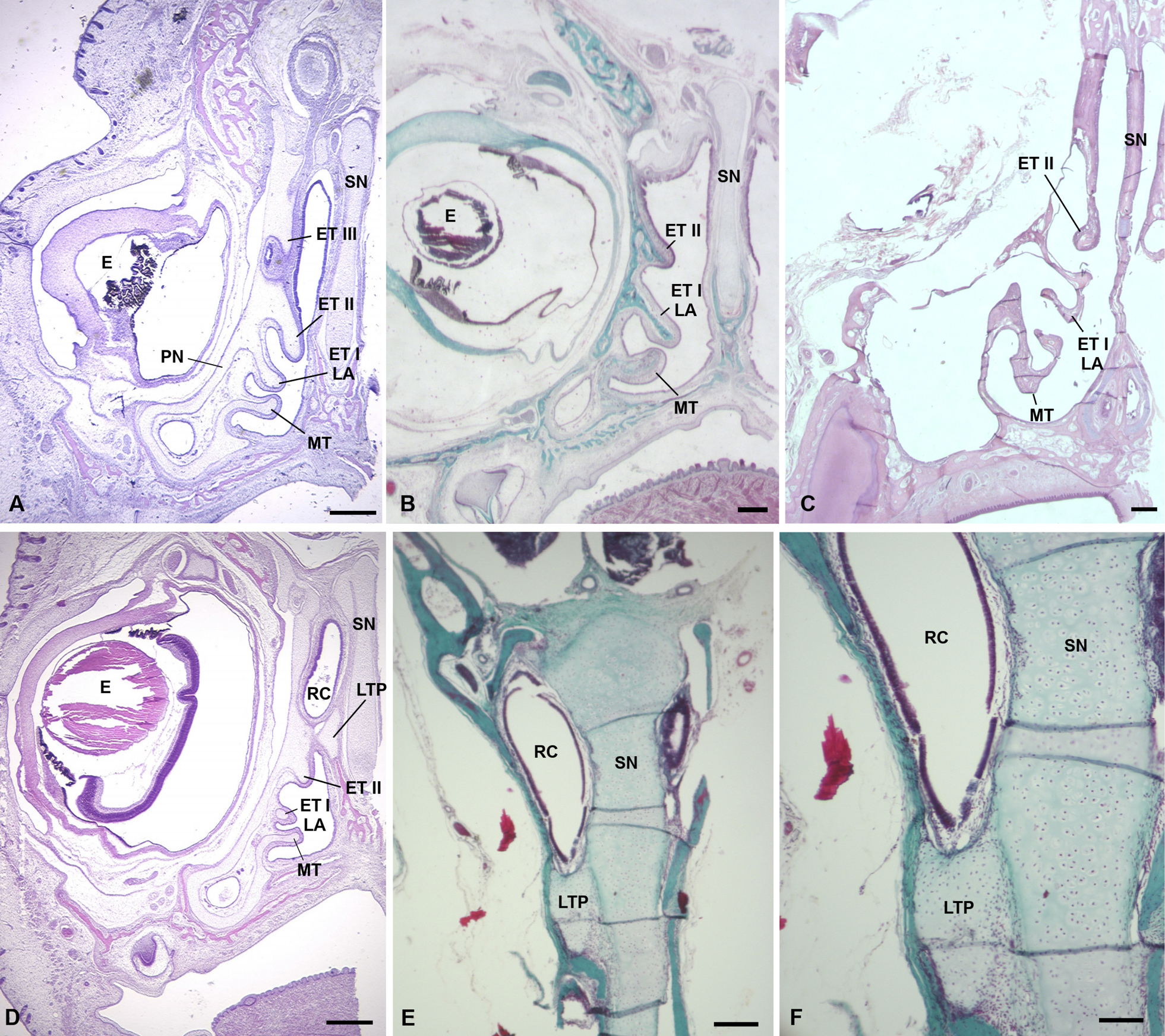
Histological serial sections of the nasal capsule in tamarins (Saguinus spp.) in coronal view (rostral to caudal). A Mid-fetal S. geoffroyi (SG10) showing an entirely cartilaginous nasal capsule, in which three ethmoturbinals (ET I, II, and III) are visible. B In a newborn S. geoffroyi (SG3), only two ETs are seen, and both are at least partly ossified. C In an older infant S. geoffroyi (MM105), two ETs are present and are projected to a greater degree (C). Figs D through F are sectioned through the cupular recess (RC), which is supported ventrally by the posterior transverse lamina (LTP). In the fetus (D) the LTP ends posteriorly as an isolated cartilaginous process. E, F In a juvenile S. midas (Smidas), the LTP remains partially cartilaginous. Abbreviations: E, eye; LA, lamina anterior of ET I; MT, maxilloturbinal; PN, paries nasi; SN, septum nasi. Scale bars: 0.4 mm (A, D); 0.5 mm (B, C); 0.2 mm (E); 0.1 mm (F). |