|
||
|
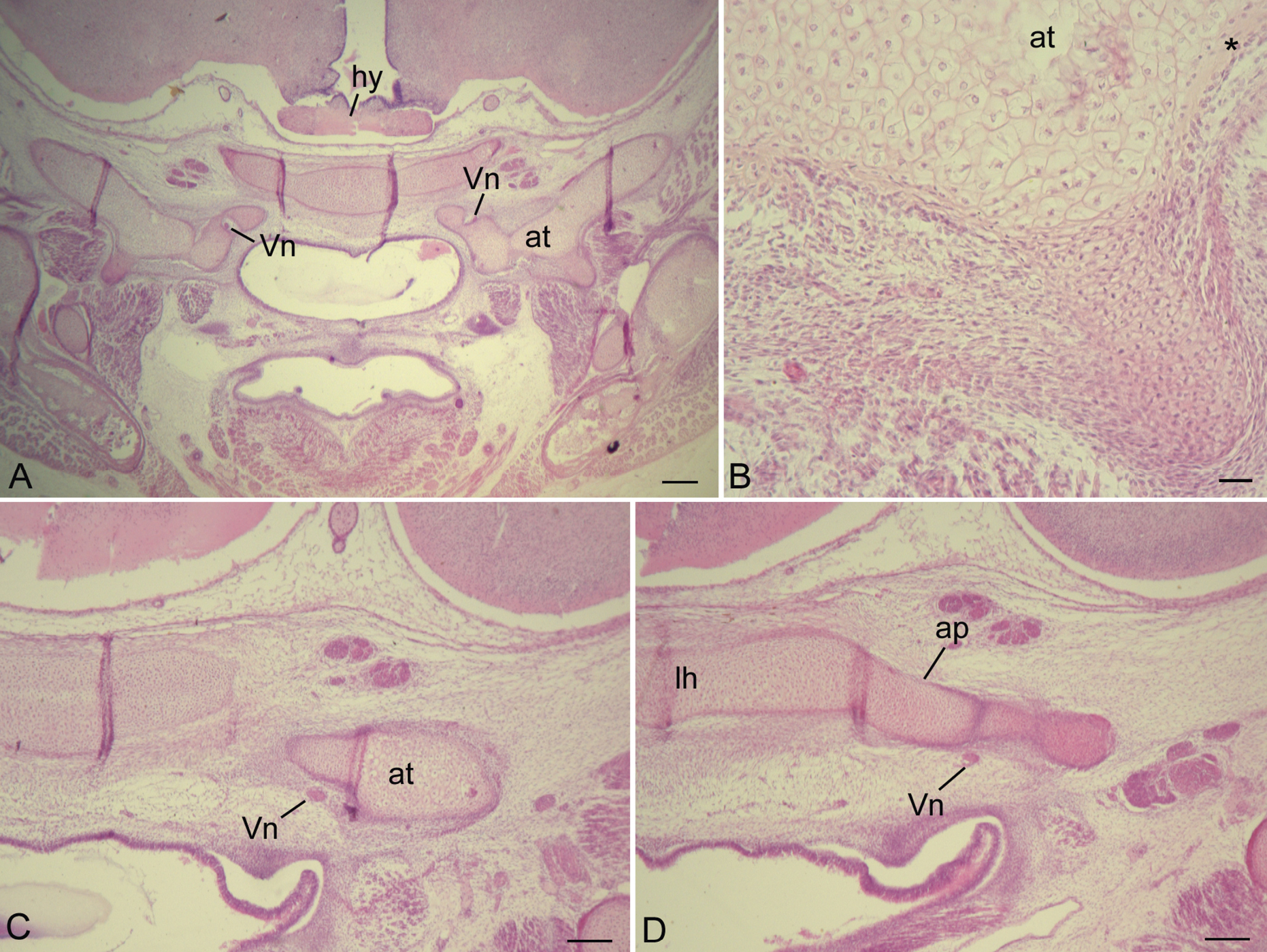
Histology of prenatal Nycticebus coucang sphenoid bone, as seen in coronal sections. A) Anterior aspect of developing sphenoid region, where the Vidian nerve (vn) passes medially and superiorly to ala temporalis (at). B) Magnified view of AT, same section as plate a, revealing densely-packed chondrocytes, and newly formed perichondrial bone (*). C) A more posterior section shows the alatemporalis to be much smaller, and further posteriorly (D) it connects to the alar process (ap) of the lamina hypophyseos (lh); here, the Vidian nerve passes inferiorly. Further abbreviations: hy, hypophysis. Scale bars: A, 250 µm; B, 30 µm; C, D, 150 µm. |