|
||
|
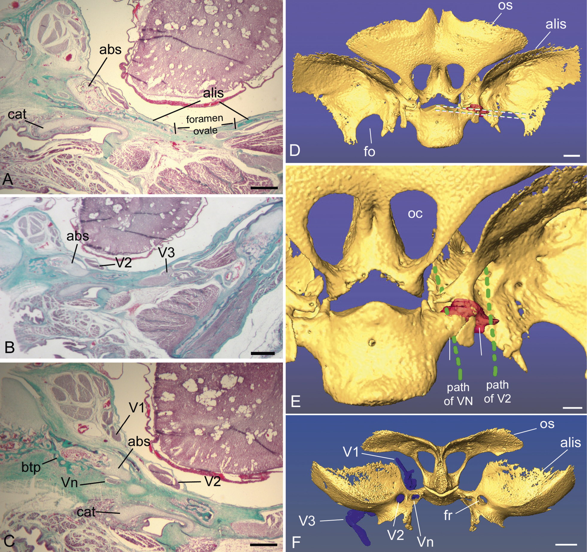
Histology of the alibasisphenoidal synchondrosis (abs) in a perinatal Saimiri boliviensis, shown in coronal sections. A–C) anterior to posterior sections, showing that the abs connects to the alisphenoid (alis) anteriorly (A), and to the basitrabecular process (btp) of the basisphenoid (bs) posteriorly (C). D-F) Reconstruction of the same stillborn Saimiri boliviensis based on aligned CT and histology slices. D) Sphenoid shown in dorsal view with the cartilaginous abs shown in red. The white dashed lines indicate the levels of section A and C. E) Magnification of the same view, with the path of the maxillary (V2) and Vidian nerve (Vn) indicated by green dashed lines. F) Sphenoid, in anterior view, shows all three major trigeminal nerve branches and the course of each nerve passing through its respective foramina. Further abbreviations: cat, cartilage of auditory tube; fo, foramen ovale; os, orbitosphenoid; fr, foramen rotundum. oc, orbital canal; ps, presphenoid; iss, intrasphenoid synchondrosis; sof, superior orbital fissure; V1, ophthalmic division of trigeminal nerve; V3, mandibular divisions of trigeminal nerve. Scale bars: A–C, 0.5 mm; D, 1 mm; E, F, 1.5 mm. |