|
||
|
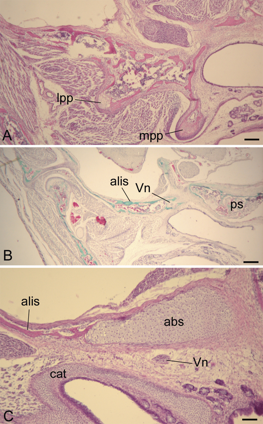
Histology of three components of sphenoid bone in fetal Loris tardigradus, as seen in the coronal plane. A) Cartilage shown at bases of medial pterygoid (mpp), lateral pterygoid plates (lpp), and the alisphenoid (alis). B) The alisphenoid projects anterior to the basisphenoids, adjacent to the presphenoid (ps). Here the Vidian nerve (Vn) can be seen near its departure from the pterygoid canal. C) The alibasisphenoidal synchondrosis (abs), with membranous expansion of the alisphenoid emanating from its lateral limit. The Vidian nerve is inferior to the abs. Further abbreviations: cat, cartilage of the auditory tube. Stains: a, c, hematoxylin and eosin; b, Gomori trichrome. Scale bars: A, 150 µm; B, 300 µm; C, 75 µm. |