|
||
|
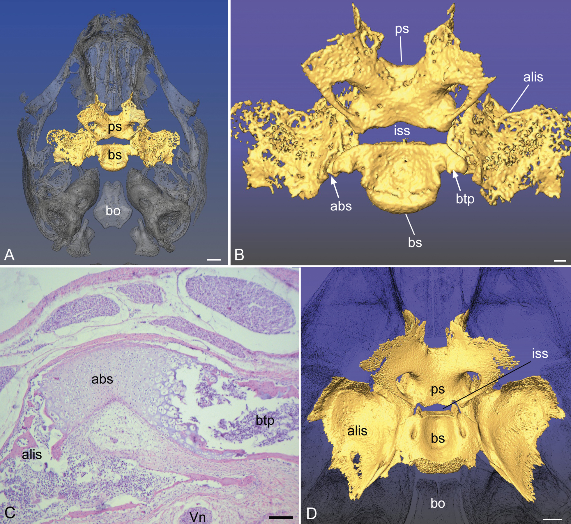
Fetal (A, B, C) compared to newborn (D) Lemur catta. A) The sphenoid bones are in the dorsal view, with ghosted skull for context. B) A more magnified view of the fetus reveals the basitrabecular process (btp) of the basisphenoids, positioned adjacent to the alibasisphenoidal synchondrosis (abs). The gap at the abs is difficult to discern osteologically, but histology (C) reveals it clearly, bridging the basiphenoid (bs) and alisphenoid. Note the Vidian nerve (Vn) approaching the posterior side of the pterygoid canal from below. D) In the newborn, histology (not shown) reveals no vestige of the cartilage of the abs. The basitrabecular process is still apparent. Note that the intrasphenoidal synchondrosis (iss) is proportionally reduced in the newborn compared to the fetus. Further abbreviations: bo, basioccipital; ps, presphenoid. Scale bars: A, 1.5 mm; B, 0.5 mm; C, 200 µm; D, 1.5 mm. |