|
||
|
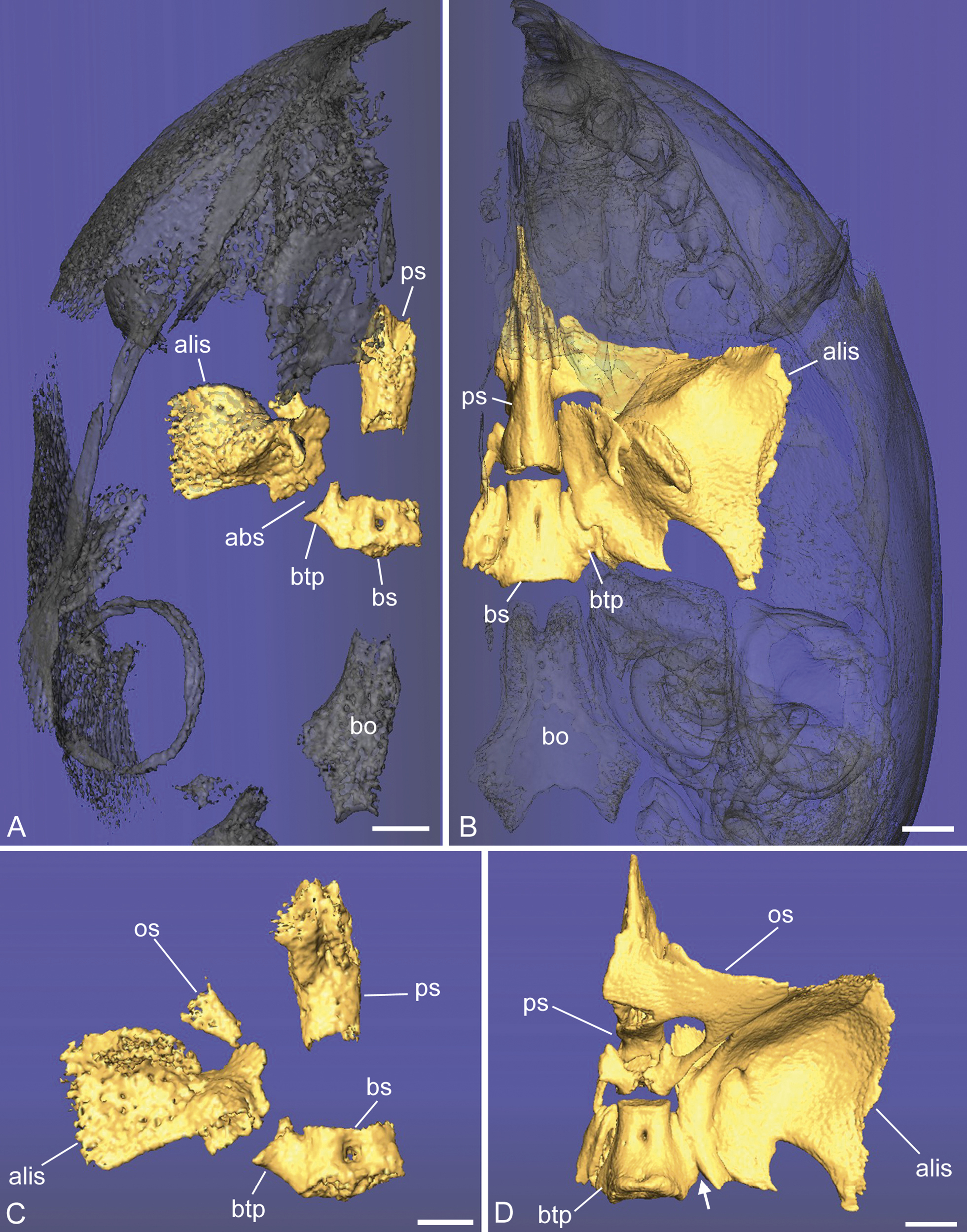
Fetal (A, C) and newborn (B, D) specimens of Saguinus geoffroyi. A, B) The sphenoid bone is shown in ventral view, with the whole skull in ghosted view. Note the marked decreased distance between the basisphenoid (bs) and presphenoid (ps). Also note that the alibasisphenoidal synchondrosis (abs), between the basisphenoid and the alisphenoid (alis), oriented laterally but also anteriorly in the fetus, appears fused or nearly so at birth. C, D) Enlarged dorsal views show the degree of ossification of the sphenoid overall. The fetal specimen has a more precociously ossified basisphenoid and alisphenoid relative to the presphenoid and orbitosphenoid (os). In the enlarged view of the fetus (C), the orbitosphenoid is shown to be in a very early point in ossification; the ossified element is the inferior (metoptic) root. Also note the position of the basitrabecular process (btp), located at the posterior margin of the abs. This landmark can also be seen in the newborn (D, arrows), but the synchondrosis is greatly reduced at birth. Further abbreviations: bo, basioccipital. Scale bars: A, 1 mm; B, 1.5.mm; C, 750 µm; D, 1.5 mm. |