|
||
|
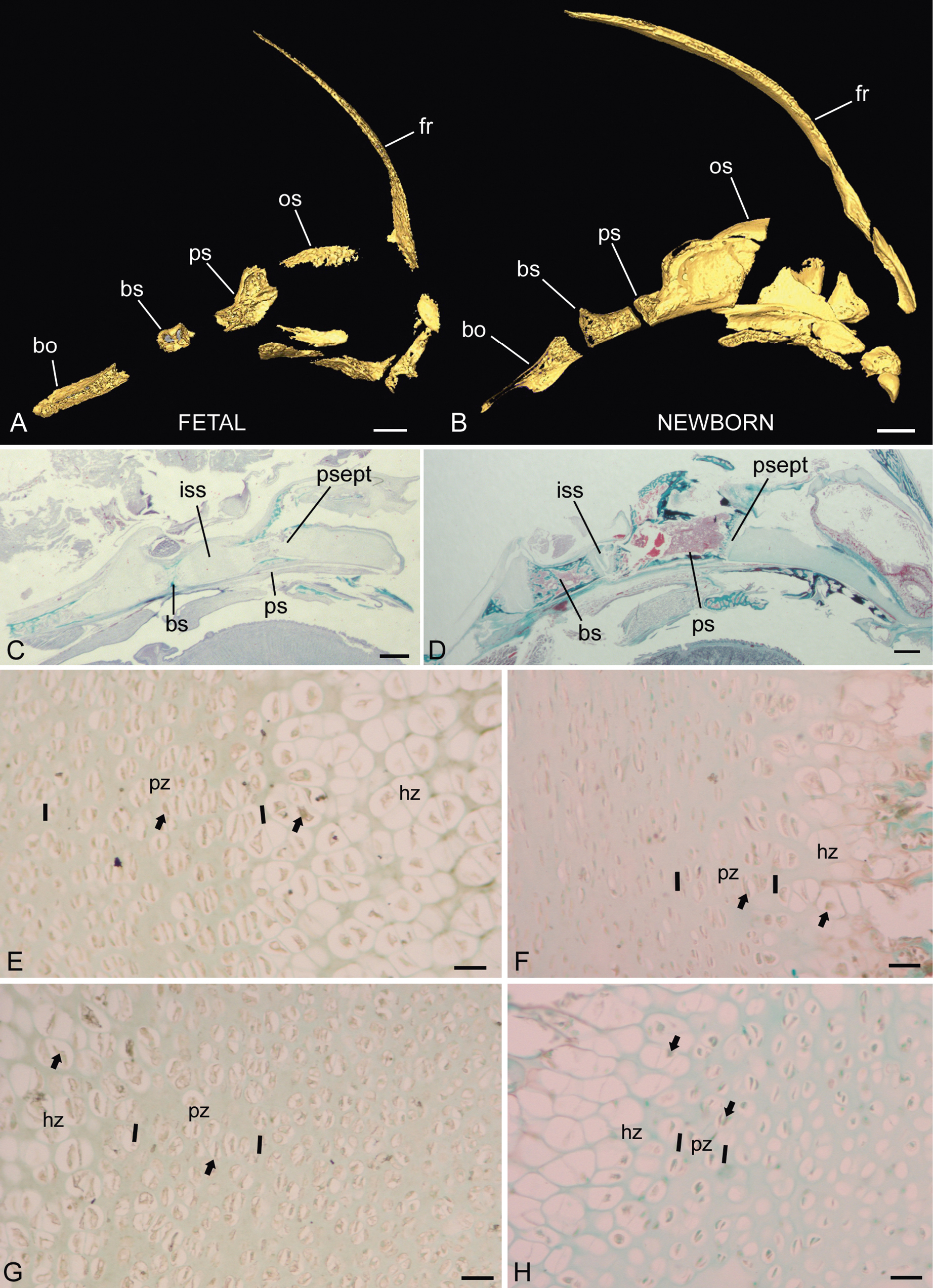
Fetal (left side) and newborn (right side) midline synchondroses in Saguinus spp. A, B) Midline segments of Saguinus geoffroyi crania are shown at the two ages revealing the decreased space between the presphenoid (ps) and basisphenoid (bs) bones in the newborn (B) compared to the fetus (A). C, D) low magnification views of the midline synchondroses in the fetus (C, same specimen as A) and a newborn (D, S. oedipus used instead due to better preservation); both stained with Gomori trichrome procedure. Indicated are the intrasphenoidal (iss) and presphenoseptal (psept) synchondroses. Below, higher magnifications of the iss (E, F) and PSept (G, H) at the same ages, prepared using PCNA immunohistochemistry, with fast green counterstain. Since all higher magnification images are at the same magnification, it is clear that the absolute length of the zones of proliferating (pz) and hypertrophic (hz) chondrocytes is reduced in the newborn compared to the fetus. Further abbreviations: fr, frontal; os, orbitosphenoid; bo, basioccipital. Scale bars: A, 1 mm; B, 2 mm; C, D, 1 mm; E–H, 30 µm. |