|
||
|
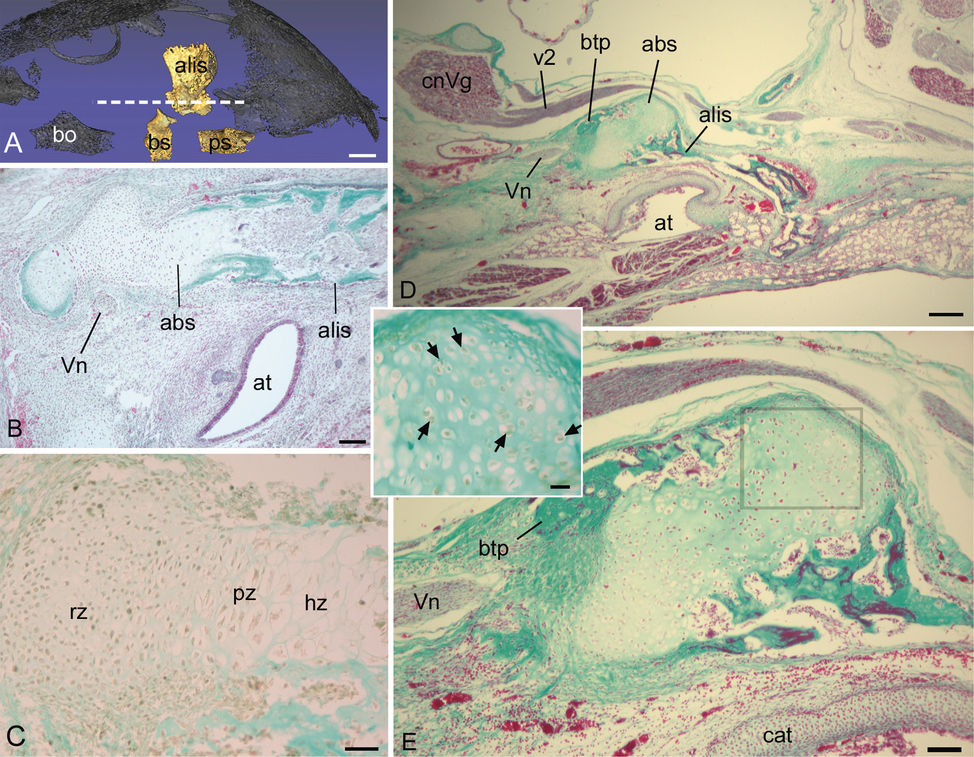
The alibasisphenoidal synchondrosis (abs) in late fetal Saguinus geoffroyi (A–C) and newborn Saguinus oedipus (D, E). A) The approximate level of the sagittal histological section of the fetal sphenoid bone is shown as a dashed line over the basal view of the skull (sphenoid emphasized, remainder of skull ghosted in black). B) a parasagittal section revealing an elongated abs. C) a section near the one shown in B shown at higher magnification, prepared using immunohistochemistry to PCNA, counterstained with fast green. Note chondrocytes in the reserve (rz) and proliferating zones (pz) are PCNA+, but most chondrocytes in the hypertrophic zone (hz) are PCNA-negative. D, E) low and higher magnifications of abs in newborn S. oedipus. Note the growth is now re-directed slightly inferiorly from the basitrabecular process (btp) of the basisphenoid bone, and the abs is proportionately shorter in anteroposterior breadth. Inset: A preparation of a nearby section to that in e (see box for specific location), made using immunohistochemistry to PCNA, counterstained with fast green. Multiple chondrocytes are PCNA + (arrows), but few chondrocytes are organized into columns. Note the close relationship of abs to the Vidian nerve (Vn). Further abbreviations: alis, alisphenoid; at, auditory tube; bo, basioccipital; bs, basisphenoid; cat, auditory tube cartilage; cnVg, trigeminal ganglion; ps, presphenoid. Stains: Gomori trichrome, except for inset (see above). Scale bars: A, 1mm; B, 100 µm; C, 40; D, 250 µm; E, 80 µm; inset, 20 µm. |