|
||
|
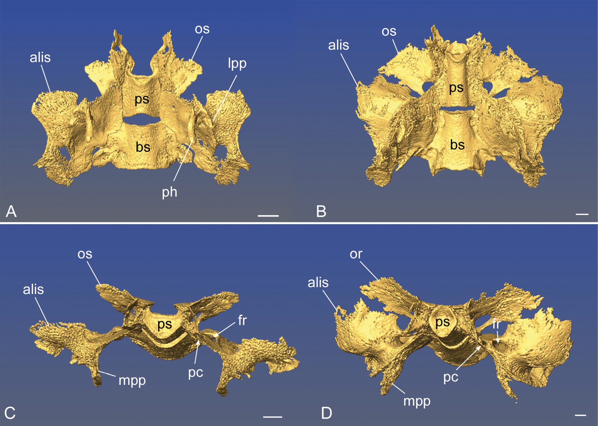
The sphenoid bone in two specimens of Varecia spp, revealing perinatal transformation of the different components of the bone. A, C) a stillborn specimen of V. rubra that was undersized compared to newborn specimens, and presumably at a late fetal stage. B, D), a 24-day-old infant V. variegata. The top row shows a ventral view, the bottom row shows an anterior view, slightly lateral to the left side. Further abbreviations: alis, alisphenoid; bs, basisphenoid; fr, foramen rotundum; mpp, medial pterygoid plate; lpp, lateral pterygoid plate; os, orbitosphenoid; ph, pterygoid hamulus; pc, pterygoid canal; ps, presphenoid. Scale bars: A, C, 1.5 mm; B, D, 1 mm. |