|
||
|
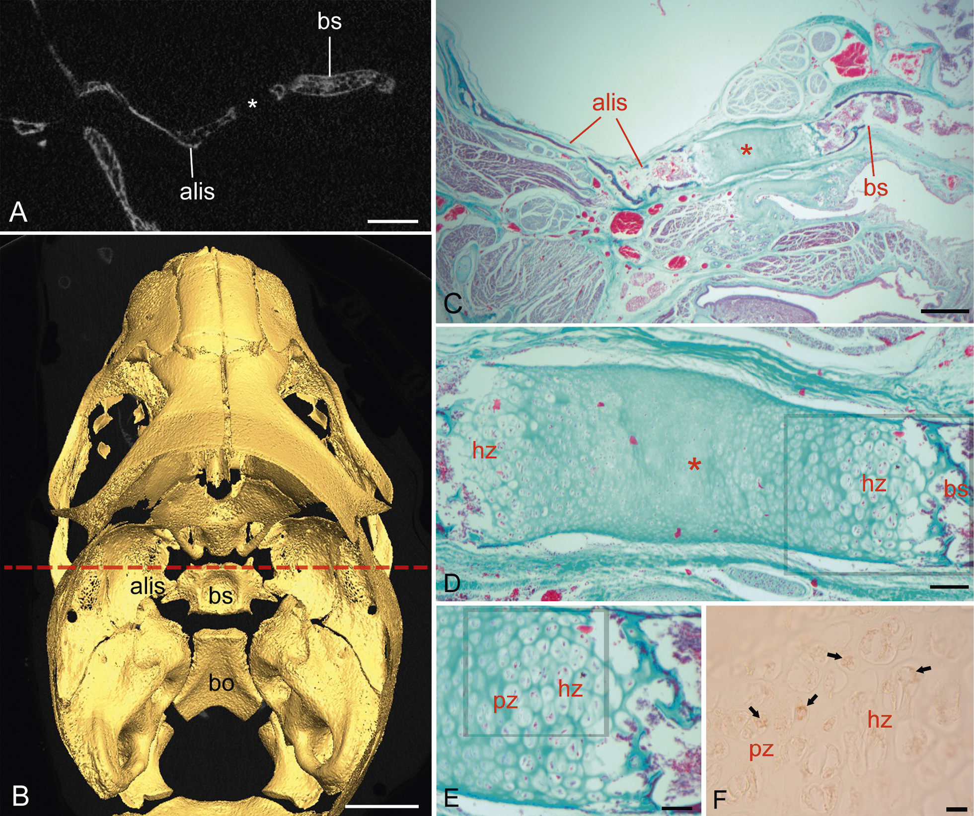
Late fetal cranium of Otolemur crassicaudatus, showing the synchondrosis (*) between the alisphenoid (alis) and basisphenoid (bs). The slice plane in CT at top left (A) is indicated by a red dashed line through the endocranial view of the skull (B). C) Histology from a twin sibling, closely matching the CT slice plane. D) A magnified view shows this is a bipolar synchondrosis, with a zone of hypertrophic chondrocytes (hz) adjacent to the alisphenoid and basisphenoid. E) A higher magnification view, enlarged from boxed area in D reveals rows of proliferating chondrocytes (pz) adjacent the hypertrophic zone. F) A higher magnification view in a nearby section is prepared with PCNA immunohistochemistry. Note PCNA-reactive chondrocytes (arrows), which are strongly reactive in proliferating chondrocytes and moderately in hypertrophic chondrocytes (box in (E) shows the approximate location of plate f). Further abbreviations: bo, basioccipital. Scale bars: A, 1.5 mm; B, 4 mm; C, 0.5 mm; D, 100 µm; E, 50 µm; f, 10 µm. |