|
||
|
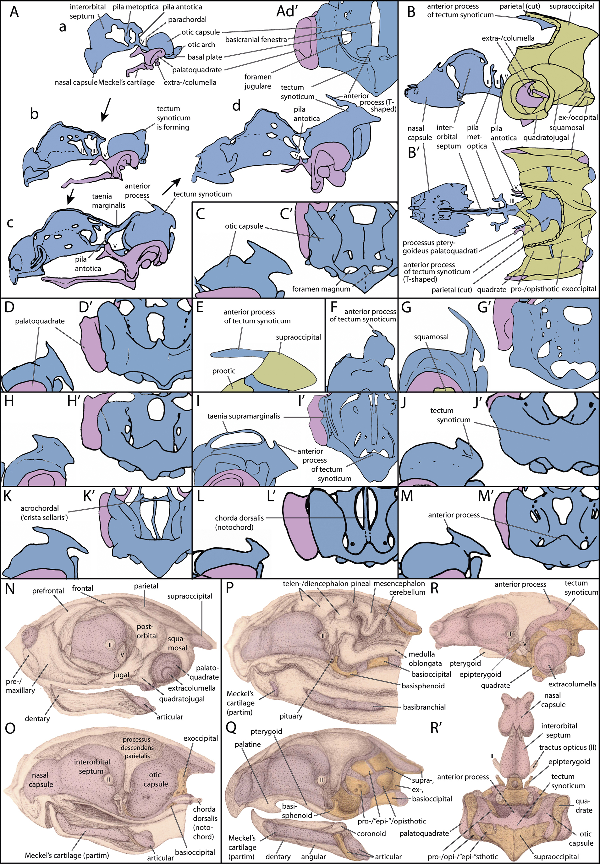
Late embryonic diversity of the anterior (tectal) process in different turtle species in lateral (normal letter) and dorsal (letter with ‘) views (redrawn from cited references). In Ad’ and C–L’, only parts of the chondrocrania are shown with the left otic capsule for orientation. Earlier stages are shown in C, D, J, M), post-hatching specimens are shown in P–R’. A) four developmental stages of Caretta caretta (Kuratani 1999), embryos with a carapace length (CL) of a) 9.2–9.7 mm, b) 11.6–12.6 mm, c) 13.1–14.1 mm, d) > 16.6 mm; B) hatchling of Podocnemis unifilis (Sheil and Zaharewicz 2014) with different ossification in the braincase (dermal bones not shown, only parietal cut); C) Apalone spinifera (Sheil 2003); D) Chelydra serpentina (Sheil & Greenbaum 2005); E) Chrysemys marginata (Shaner 1926); F) Emydura subglobosa (Werneburg & Yaryhin 2019), G) Emys orbicularis (Kunkel 1912); H) Eretmochelys imbricata (Sheil 2013); I) Eretmochelys imbricata (Fuchs 1915); J) Macrochelys temminckii (Sheil 2005); K) Pelodiscus sinensis (Sánchez-Villagra et al. 2009); L) Phrynops hillarii (Bona & Alcalde 2009). M) Trachemys scripta (Tulenko & Sheil 2007); N–R’) Chelonia mydas (Parker 1880); N–O) embryo two-thirds ripe (head length: ~ 14.8 mm) with N) all bones in lateral view and O) a median sagittal section; P–R’) ripe young (head length: ~ 23.3 mm) in P) median section, Q) more lateral sagittal section, R) a similar section with less dermal bones shown, and R’) a dorsal view on the chondro-/neurocranium. Coloration in A–L follows Werneburg & Maier (2019: fig. 1): Blue, chondrocranium (cartilage); purple, viscerocranium (cartilage); green, bone (endochondral ossification). Images not to scale. |