|
||
|
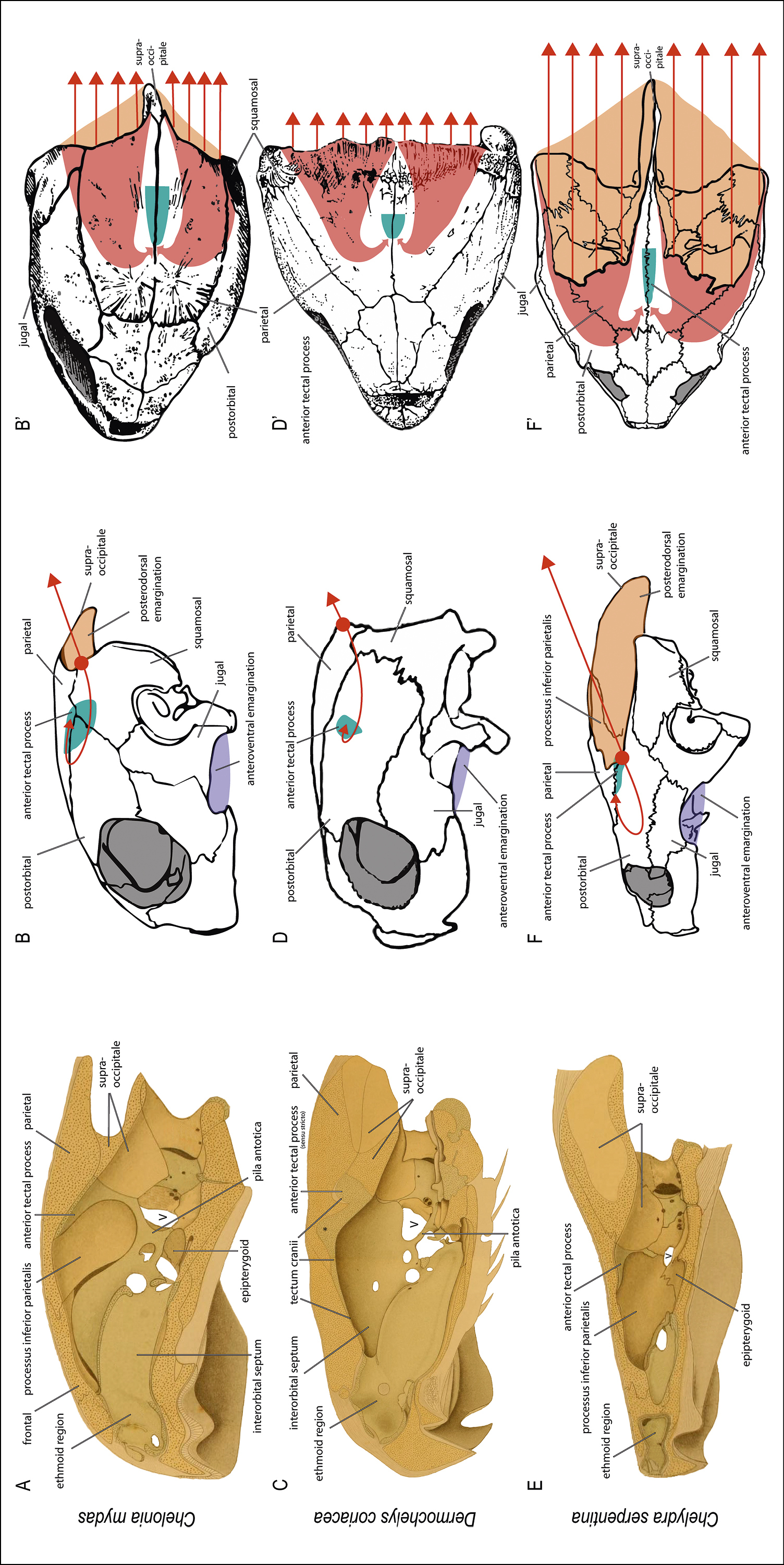
Illustration of the cartilaginous anterior process of the supraoccipital and hypothesis on the force transmissions in the skull. Note the different degrees of ossification in the skull in A, C, and E (note: these images are not exactly on the midline). In D. coriacea (C), tectum cranii is fully cartilaginous and continuous with the interorbital septum and – like in Chelo. mydas (A) – with the orbitotemporal region. In Chely. serpentina (E), only the cartilaginous process and few remainders of the cartilaginous cranium are present. The schematic illustrations show the approximate position of the anterior tectal process (blue). Bended arrows indicate suggested force transmissions in the skull: in lateral skull view (B, D, F), they are indicated by a thin bended arrow due to visualization reasons; in dorsal views (B’, D’, F’), those arrows correspond to the broad red bended arrows in the skull. The straight arrows indicate pulling neck forces, the relative degree of which are indicated by the depth of the posterodorsal emarginations. It is hypothesized that these forces in the skull are absorbed by the anterior tectal process, which has an altering relative length and position in each species. * in C indicates the spatial shift of the skull roof vs. the braincase resulting in a dorsal excavation, which is here filled with cartilage of tectum cranii – more medial, there is a space for the dorsally erected pineal gland (figs 192, 201 and 202 in Wyneken, 2001, and discussed by Paulina-Carabajal et al. 2013, 2017). Images modified after Nick (1912) (A, C, E), after Gaffney (1979) (B, F, F’), after Zangerl (1948) (D), and after Wyneken (2001) (B’, D’). |