|
||
|
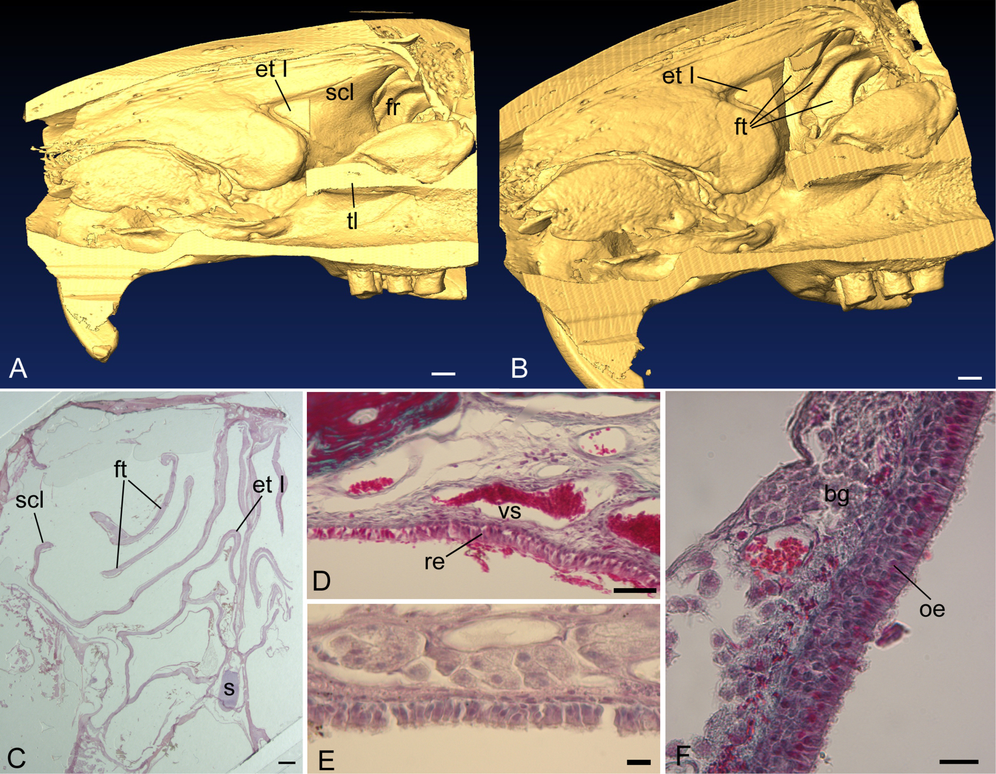
A), B), medial and rostromedial perspectives of the right nasal fossa in an adult agouti (Dasyprocta leporina). In A, the caudal part of the first ethmoturbinal (etI), the entire second and third ethmoturbinals, as well as their root lamella are virtually dissected away to reveal the semicircular lamina (scl). Caudal to the scl is the opening into the frontal recess (fr). In B, the scl is also dissected away to reveal three large frontoturbinals (ft) within the frontal recess. C–F) Histological sections in the frontal recess of Dasyprocta cristata. C, a histological section through the rostral, free projections of frontoturbinals 2 and 3, also emphasized the greatly elongated and sickle-shaped scl (dashed line in A indicates a likely comparable cross-sectional level in D. leporina). At rostral levels, the scl and each ft is covered with pseudostratified, columnar ciliated epithelium (D, showing scl) or simple cuboidal/columnar, ciliated epithelium (e, showing ft2). F) ft3 has the most rostrally projecting olfactory epithelium (oe). bg, Bowman’s glands. re, respiratory epithelium; s, septal cartilage; vs, venous sinus. Scale bars: A, 3 mm; B, 2.5 mm; C, 1 mm; D, 50 µm; E, 10 µm; F, 20 µm. |