|
||
|
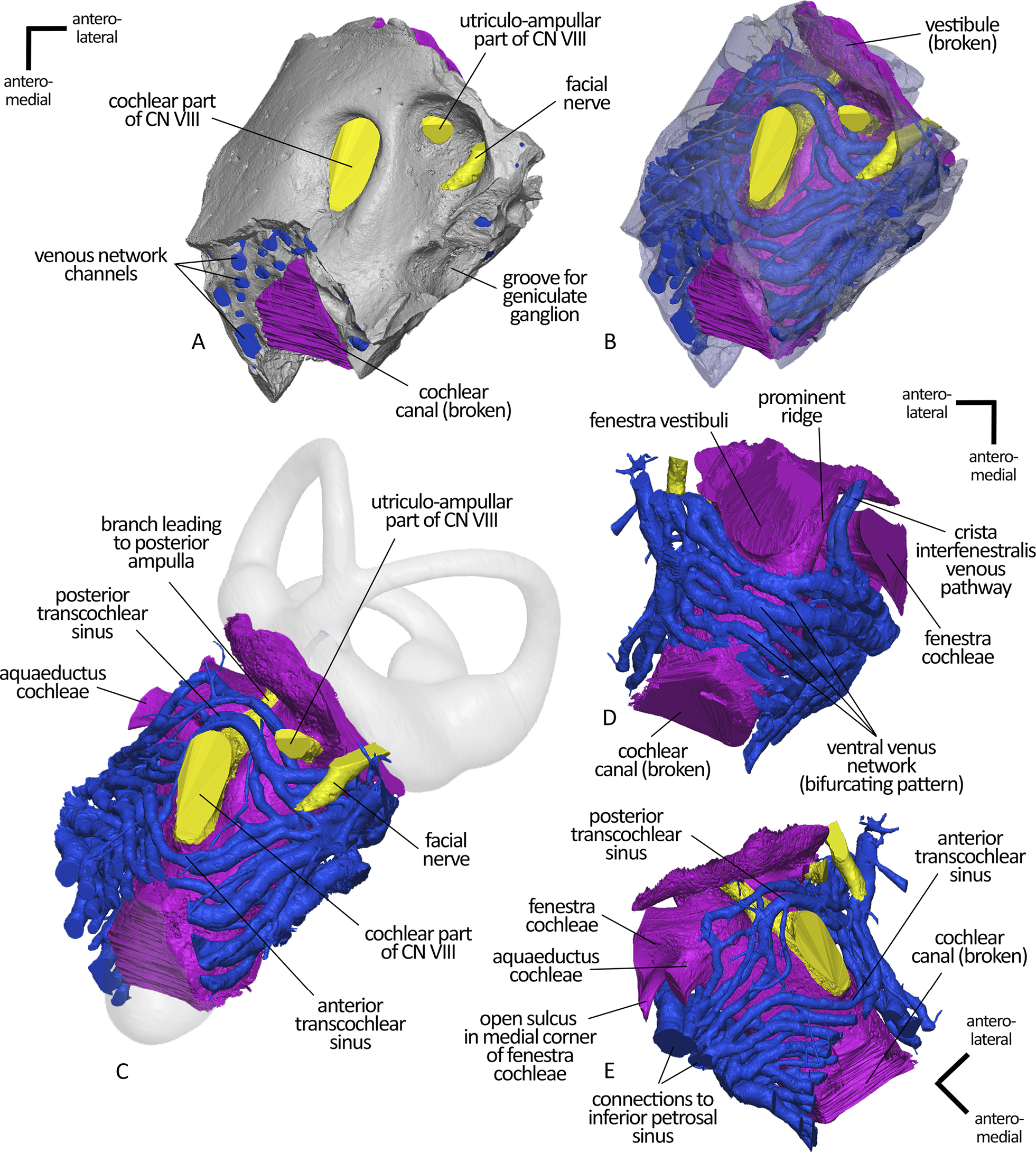
3D reconstruction of soft tissue parts of the Teete petrosal. A) Dorsal (endocranial) view of the internal acoustic meatus of the petrosal with reconstructed soft tissue parts of the cochlear endocast (pink), venous pathways (blue), and innervation areas (yellow). B) Same view as A with translucent bone parts to illustrate the high degree of vascularity of the circum-promontorial region. C) Same view as A and B, reconstructed soft tissue parts (cochlear endocast pink, venous pathways blue, innervation areas yellow) enclosed in the petrosal bone with indicated missing parts of the inner ear (vestibular region and cochlear apex in shaded gray). D) Ventral view of the bifurcating venous pathways. E) Dorsomedial view of the reconstructed soft tissue parts (cochlear endocast pink, venous pathways blue, innervation areas yellow). |