|
||
|
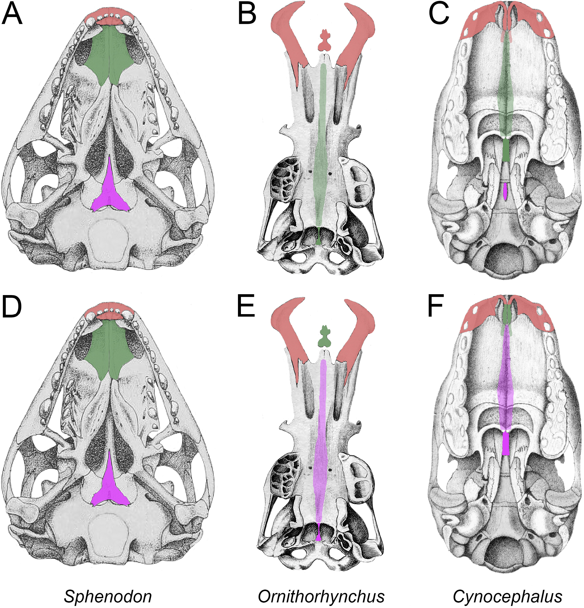
Crania in ventral view. A, D, Sphenodon punctatus (modified from Howes and Swinnerton 1901: plate IV, fig. 6); B, E, Ornithorhynchus anatinus (modified from Meckel 1826: table IV, fig. I); C, F, Cynocephalus volans (modified from Parker 1885b: plate 37, fig. 6). In A–C, bones are color coded following the hypothesis that the sauropsid vomer (prevomer of Broom 1895) = the mammalian vomer [green], the platypus os paradoxum = the therian medial palatine process of the premaxilla [red], and the sauropsid parasphenoid = the therian parasphenoid [purple]. In D-F, the bones are color coded following the hypothesis that the sauropsid vomer = the platypus os paradoxum = the therian medial palatine process of the premaxilla [green] and the sauropsid parasphenoid = the mammalian vomer [purple]. Parasphenoid [purple in C] is removed in F to make the morphology congruent with the hypothesis. The parts of bones hidden by the palate in B, C, E, and F are indicated by semi-transparency. |