|
||
|
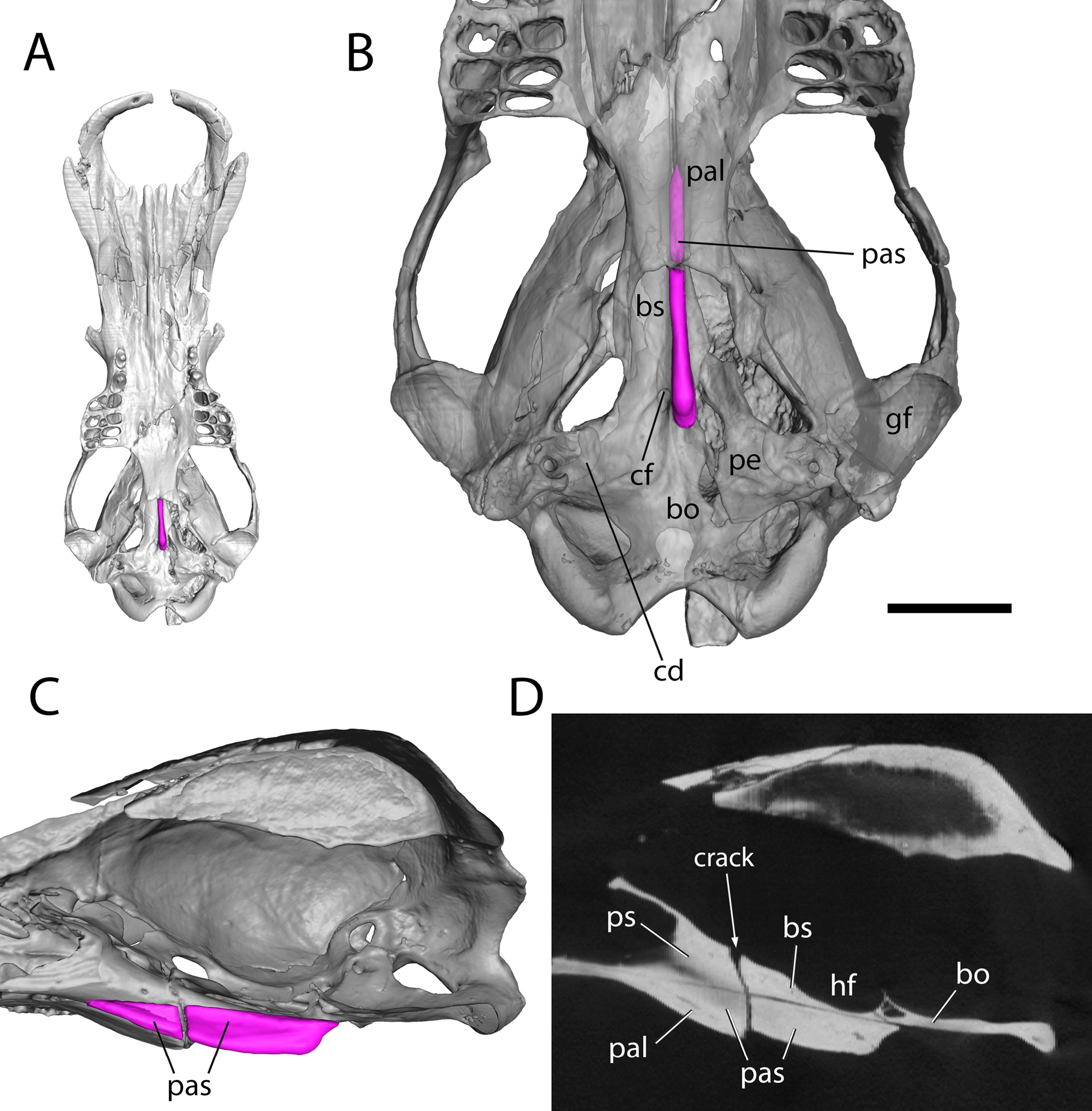
Obdurodon dicksoni, QM F20568, cranium. A–C, bone isosurfaces derived from CT scans. A, cranium in ventral view; B, semi-transparent posterior cranium in ventral view showing extension of parasphenoid dorsal to palatine bones; and C, right “half” of posterior cranium in medial view, sectioned to the left of the midline. D, CT slice in parasagittal plane corresponding to C. Scale bar for B-D is 10 mm. Pre- and basisphenoid are fused but identified based on their positions. Abbreviations: bo, basioccipital; bs, basisphenoid; cd, cochlear duct; cf, carotid foramen; gf, glenoid fossa; hf, hypophyseal fossa; pal, palatine; pas, parasphenoid; pe, petrosal; ps, presphenoid. |