|
||
|
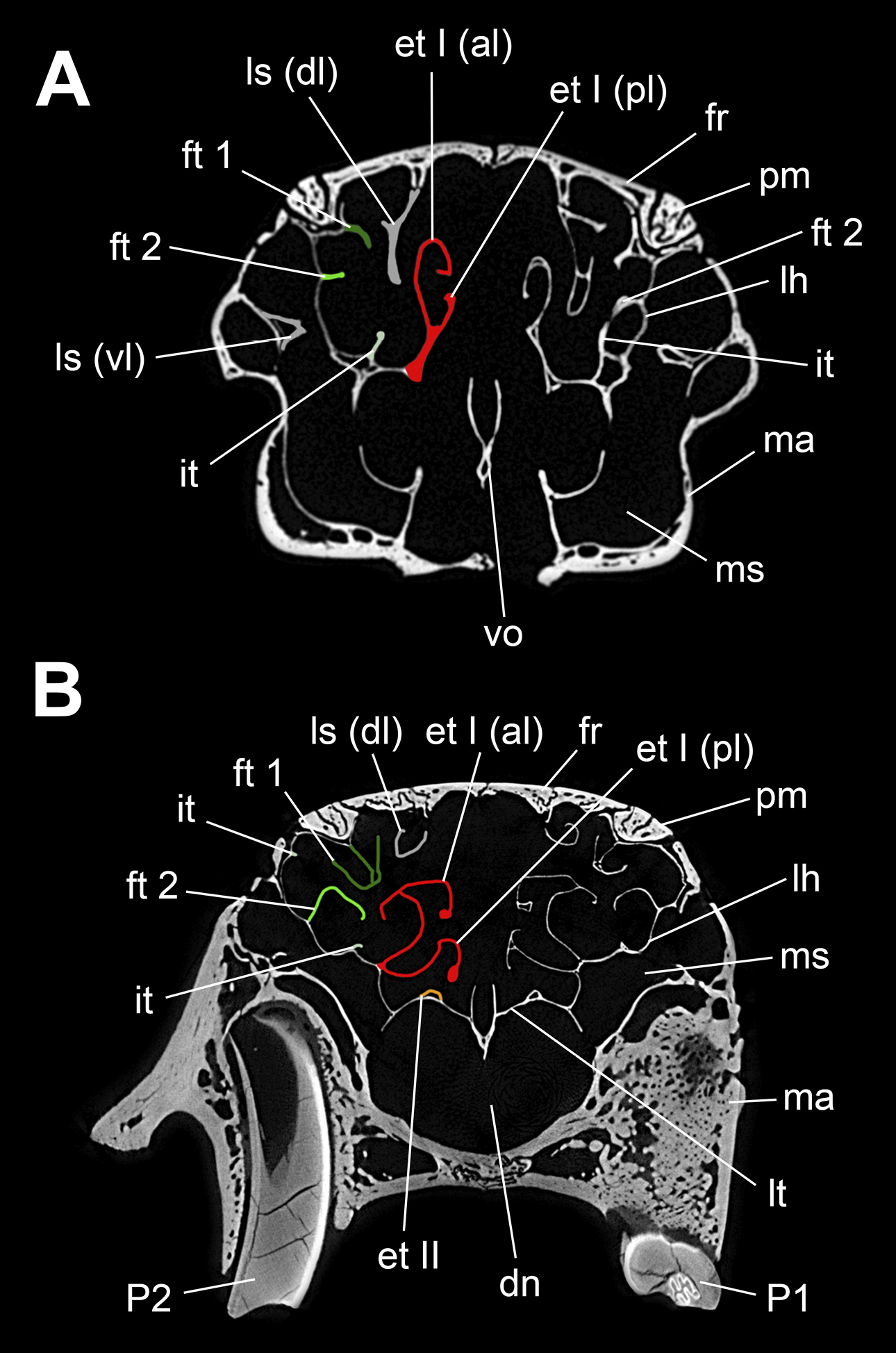
Transversal µCT images through the nasal cavity of Pentalagus furnessi. A specimen M 12940, B specimen M 12939. Note the differences in topography of the additional interturbinal between frontoturbinal 2 and ethmoturbinal I. In specimen M 12940 anteriorly this interturbinal and frontoturbinal 2 have a common origin and the former runs posteromedially to merge with ethmoturbinal I (A), whereas in specimen M 12939 the additional interturbinal is a very low ridge on the lamina horizontalis (B). Colour code of the right turbinal skeleton refers to Fig. 2. Abbreviations: al, anterior lamella; dl, dorsal lamella; dn, ductus nasopharyngeus; et I–II, ethmoturbinal I–II; fr, frontal bone; ft1–2, frontoturbinal 1–2; it, interturbinal; lh, lamina horizontalis; ls, lamina semicircularis; lt, lamina terminalis; ma, maxillary bone; ms, maxillary sinus; P1–2, upper premolar 1–2; pl, posterior lamella; pm, premaxillary bone; vl, ventral lamella; vo, vomer. Not to scale. |