|
||
|
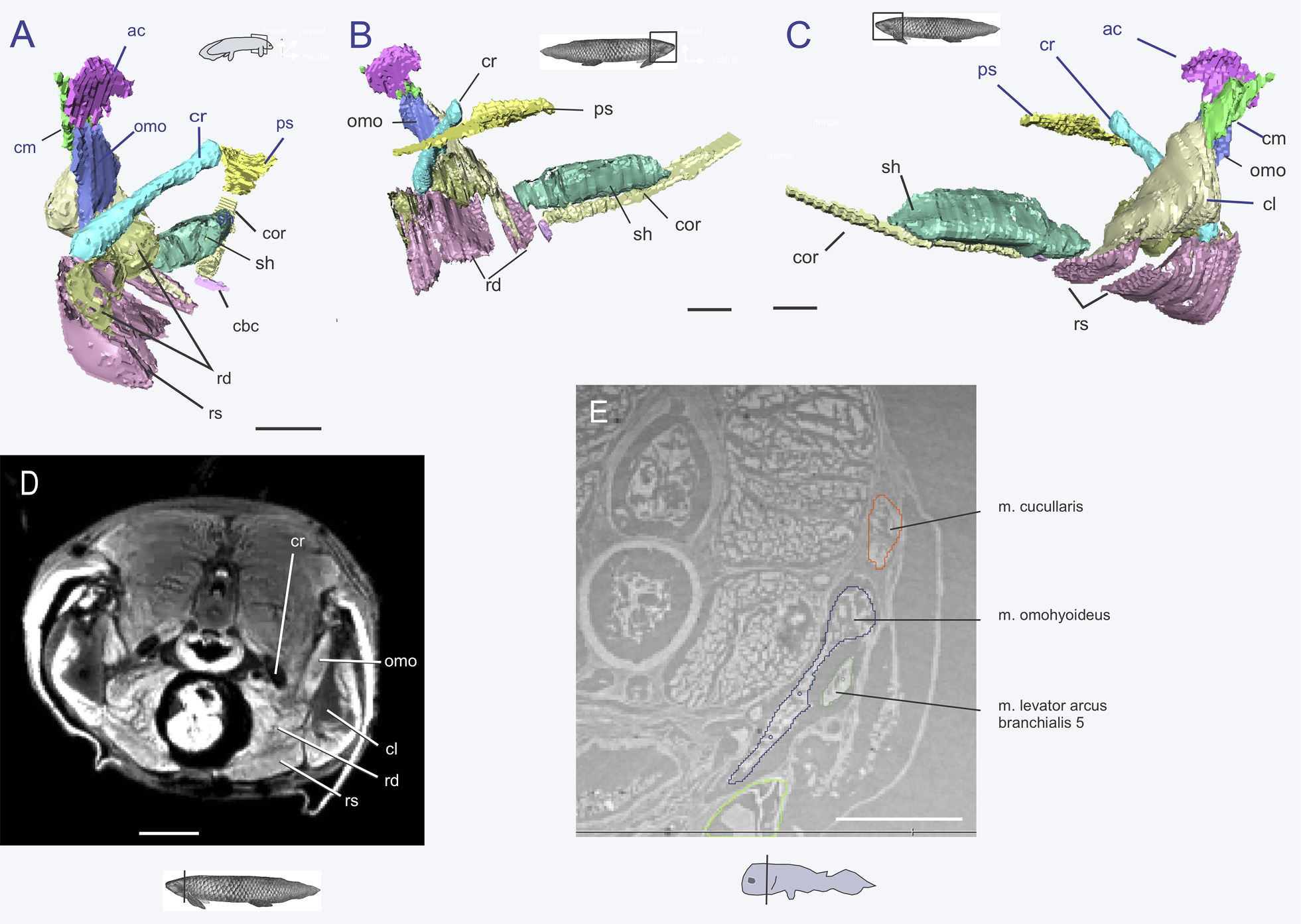
Neoceratodus forsteri , MRI scan: A–C reconstruction of muscles connected to the cranial rib in A caudomedial; B medial; and C lateral views. D slice image to demonstrate some of the structures segmented in A–C. E Latimeria chalumnae CCC 202, synchrotron tomography, slice image of segmentation to show muscle proposed as m. omohyoideus. Scale bars A–D=10mm, E=1mm. Abbreviations: ac, anocleithrum; cbc, cardiobranchial cartilage; cl, clavicle; cm, cleithrum; cor, m.coracomandibularis; cr, cranial rib; omo, m. omohyoideus; ps, parasphenoid; rs, m. rectus abdominis, superficial lamina; rd, m. rectus abdominis, deep lamina; sh, m. sternohyoideus. |