|
||
|
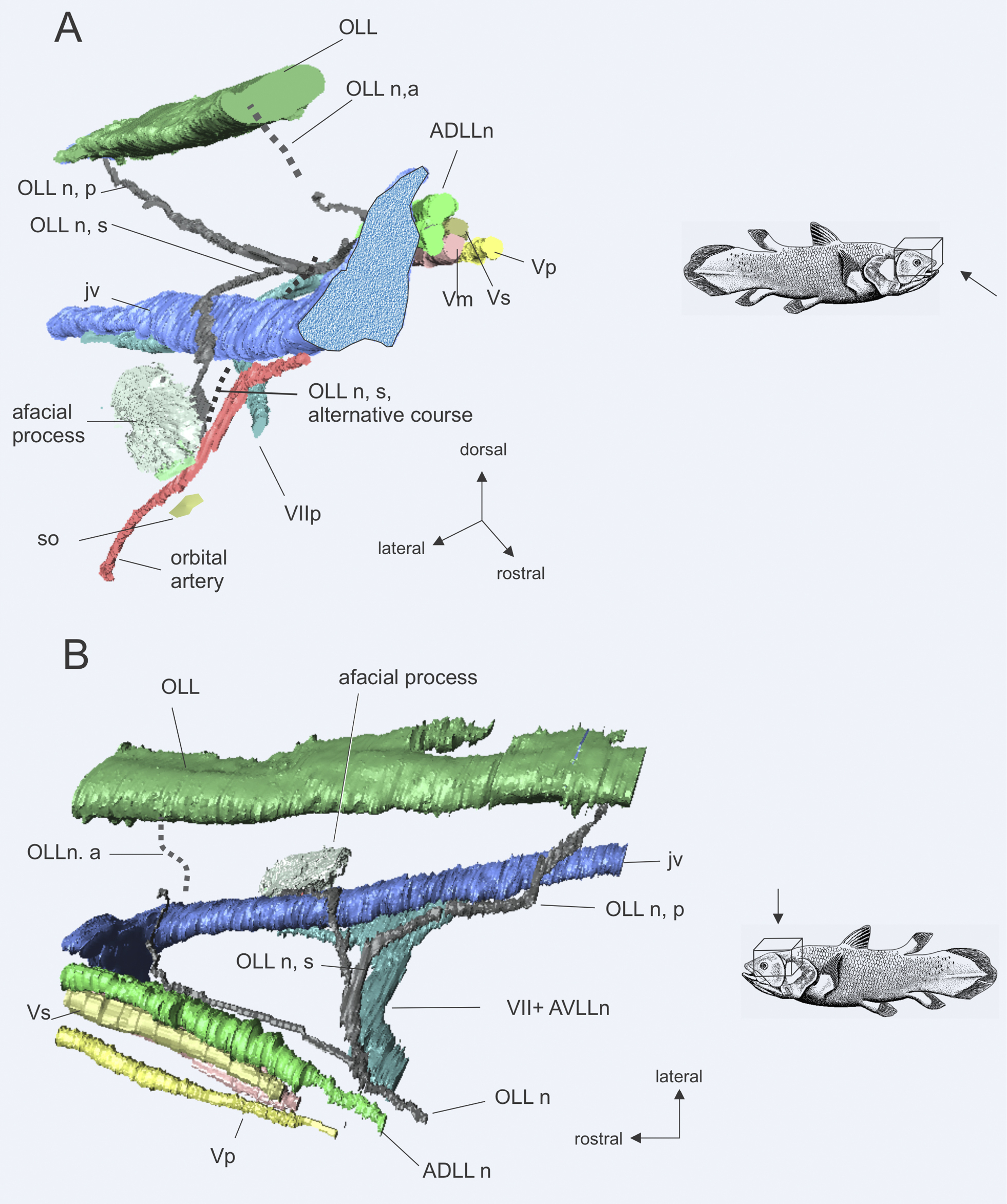
Latimeria chalumnae CCC 29.5, 3D reconstructions from synchrotron tomography, showing the otic lateral line nerve and its distribution. A anterolateral view; B dorsal view. Relationships among spiracular organ, orbital artery and palatine nerve are observed in A. Nerve branches too small to appear in the reconstruction are indicated with dashed lines. Abbreviations: OLL, otic lateral line; OLLn, otic lateral line nerve; OLLn,a, anterior ramus otic lateral line nerve; OLLn, p, posterior ramus; Olln, s, spiracular ramus of otic lateral line nerve; jv, jugular vein; so, spiracular organ; VIIp, palatine ramus of facial nerve; ADLLn, anterodorsal lateral line nerve; Vp, profundus ramus of Vth cranial nerve; Vs, sensory ramus of V nerve; Vm, motor ramus of V nerve; VII+ AVLLn, facial + anteroventral lateral line nerves. Terminology after Northcutt and Bemis (1993). |