|
||
|
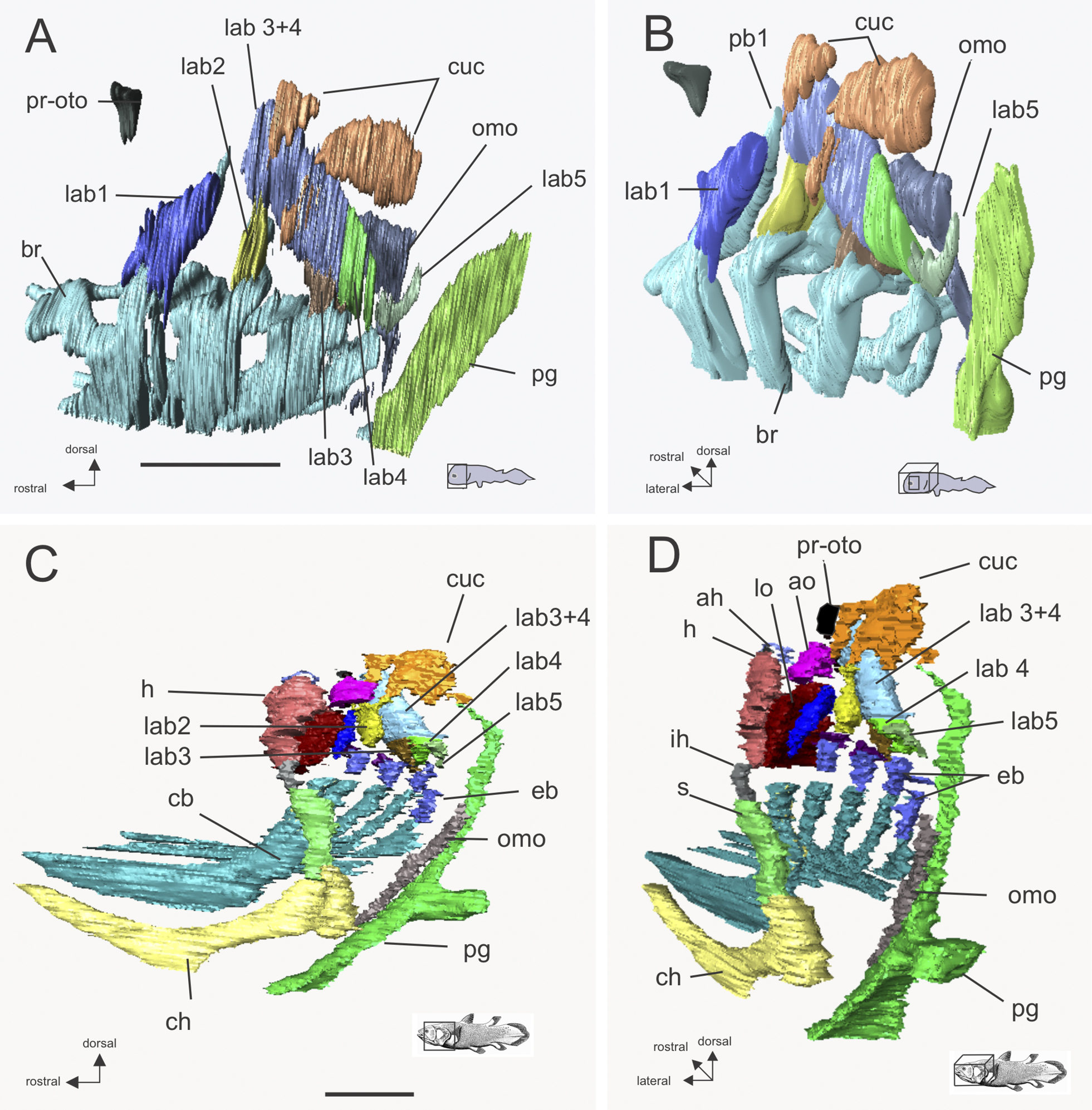
Latimeria chalumnae , 3D reconstructions: A and B: CCC 202 (fetus), synchrotron CT, and C and D, CCC 88 (adult), MRI scan, showing relationships of m. cucullaris to branchial levators and adjacent structures. Colours are similar in A, B and C, D; in C, D the epibranchial cartilages are segmented separately from the ceratobranchials, all are together in A, B. Nomenclature for the muscles of the hyomandibula and operculum in C, D as in Millot and Anthony (1958); in the scan images these three muscles form a continuous broad sheet. Abbreviations: pr-oto, location of the processus post-oticus of the braincase; br, branchial cartilages, cb, ceratobranchials, ch, ceratohyal; eb, epibranchials; pb1, suprapharyngobranchial 1; h, hyomandibula; ih, interhyale; s, sympletic; pg. pectoral girdle; cuc, m. cucullaris; omo, m. omohyoideus; lab 1–5, levator arcus branchialis 1–5 (lab 3 and 4 have a common dorsal belly which gives rise to separate muscles close to the dorsal arches); ah, m. adductor hyomandibulae; lo, m. levator operculi, ao, m. adductor operculi. Scale bars: A, B=1mm; C, D=50mm |