|
||
|
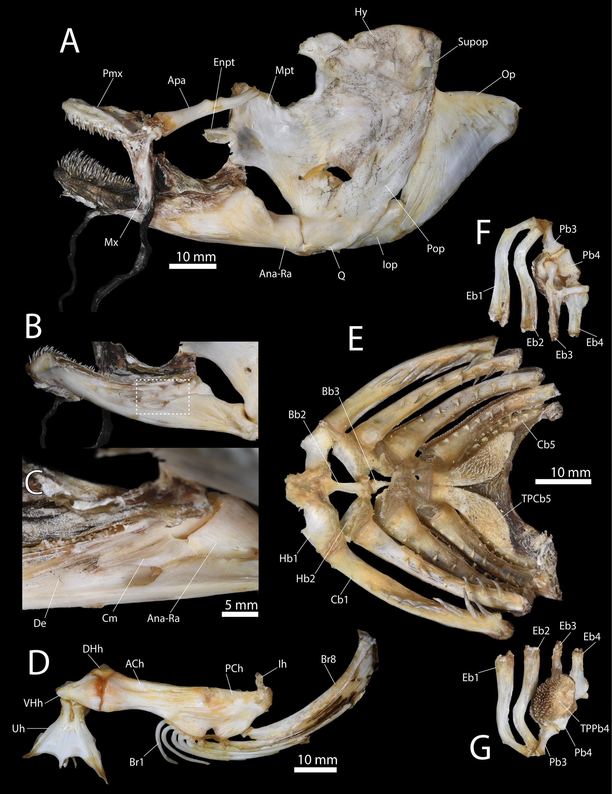
Ictalurus punctatus, specimen TCWC 20491.08, 441 mm SL (A), (B), (C), (E), and TCWC 20491.07, 436 mm SL (D). A Hyopalatine arch, jaws and opercular series in lateral view. B Lower jaw in medial view. Area outline by white box indicates location of (C). C Coronomeckelian on medial surface of the lower jaw. D Hyoid bar in lateral view. E Ventral gill arches in dorsal view. F Dorsal gill arches in dorsal view. G Dorsal gill arches in ventral view. Abbreviations: ACh, Anterior ceratohyal; Ana-Ra, Anguloarticular+retroarticular; Apa, Autopalatine; Bb, Basibranchial; Br, Branchiostegal ray; Cb, Ceratobranchial; Cm, Coronomeckelian; De, Dentary; DHh, Dorsal hypohyal; Eb, Epibranchial; Enpt, Endopterygoid; Hb, Hypobranchial; Hy, Hyomandibular; Ih, Interhyal;Iop, Interopercle; Mpt, Metapterygoid; Mx,Maxilla; Op, Opercle; Pb, Pharyngobranchial; PCh, Posterior ceratohyal; Pmx, Premaxilla; Pop, Preopercle; Q, Quadrate; Sop, Subopercle; Supop, Suprapreopercle; TPCb, Toothplate of ceratobranchial; TPPb, Toothplate of pharyngobranchial;Uh, Urohyal; VHh, Ventral hypohyal. |