|
||
|
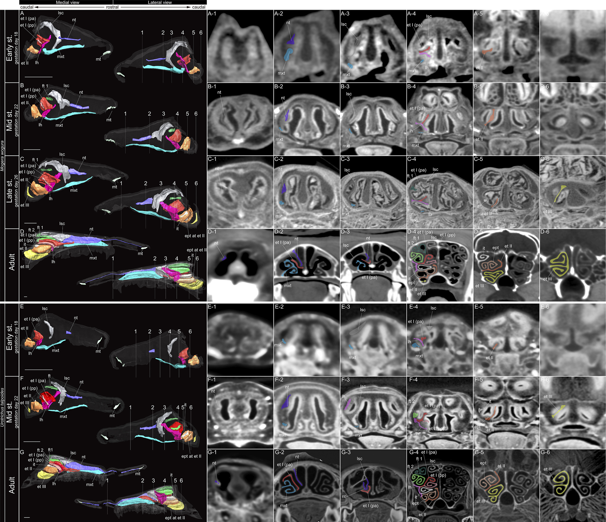
Coronal section and sagittal 3D views of µCT images of Mogera wogura and Urotrichus talpoides. A–G Show approximate location of section through the nasal capsule or nasal cavity. (A-1–6) Early stage fetus, (B-1–6) mid stage fetus, (C-1–6) late stage fetus, and (D-1–6) adult of M. wogura. (E-1–6) early stage fetus, (F-1–6) mid stage fetus, and (G-1–6) adult of U. talpoides. Scale bars: 1 mm. Abbreviations: ept = epiturbinal; et I (pa) = ethmoturbinal I pars anterior; et I (pp) = ethmoturbinal I pars posterior; et II–III = ethmoturbinal II–III; ft 1, 2= frontoturbinal 1, 2; it = interturbinal; lh = lamina horizontalis; lsc = lamina semicircularis; mt = marginoturbinal; mxt = maxilloturbinal; nt = nasoturbinal. |