|
||
|
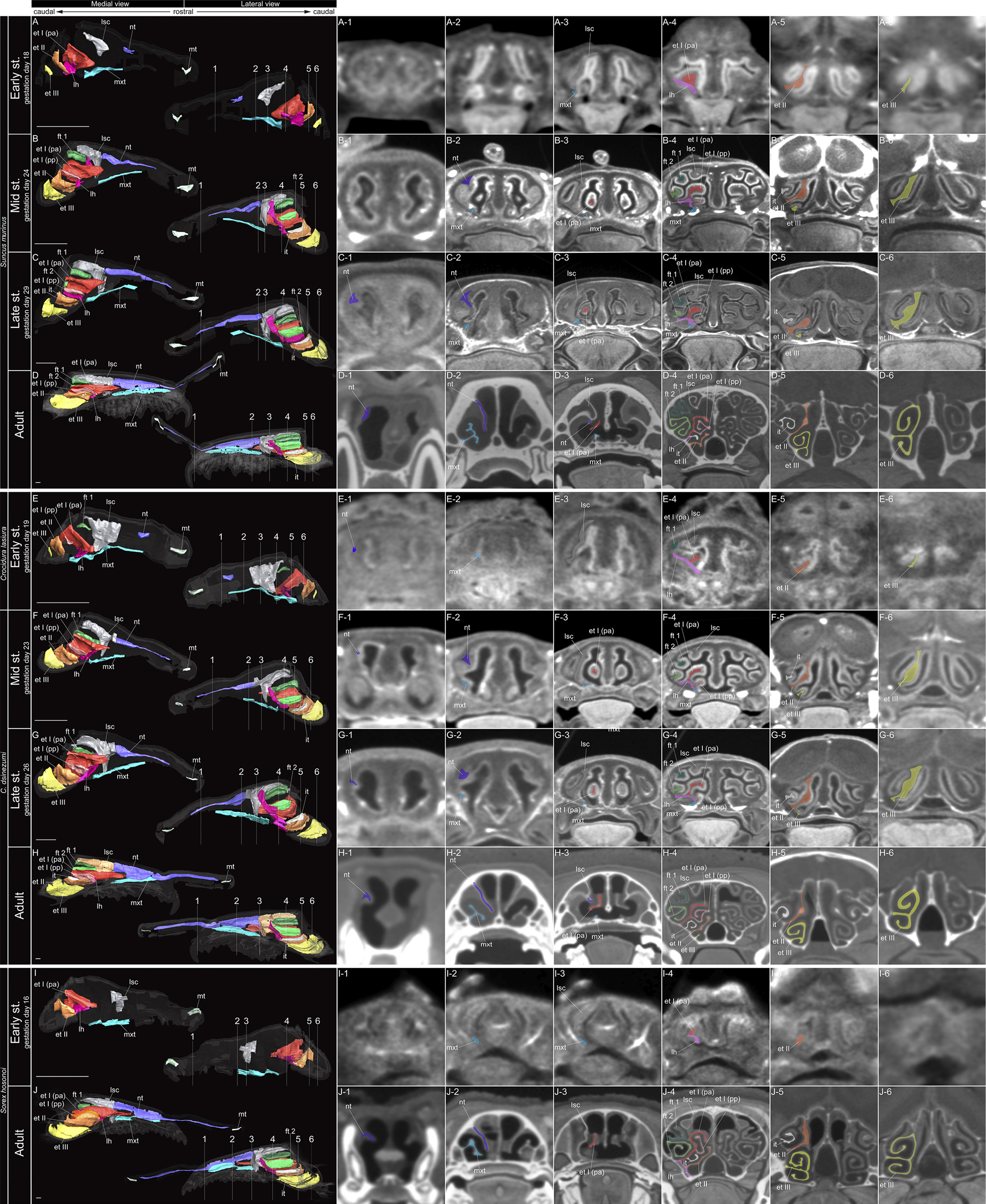
Coronal section and sagittal 3D views of µCT images of Suncus murinus, Crocidura lasiura, C. dsinezumi, and Sorex hosonoi. A–J show approximate location of section through the nasal capsule or nasal cavity. (A-1–6) Early stage fetus, (B-1–6) mid stage fetus, (C-1–6) late stage fetus, and (D-1–6) adult of S. murinus. (E-1–6) Early stage fetus of C. lasiura. (F-1–6) Mid stage fetus, (G-1–6) late stage fetus, and (H-1–6) adult of C. dsinezumi. (I-1–6) Early stage fetus, and (J-1–6) adult of S. hosonoi. Scale bars: 1 mm. Abbreviations: et I (pa) = ethmoturbinal I pars anterior; et I (pp) = ethmoturbinal I pars posterior; et II–III = ethmoturbinal II–III; ft 1, 2= frontoturbinal 1, 2; it = interturbinal; lh = lamina horizontalis; lsc = lamina semicircularis; mt = marginoturbinal; mxt = maxilloturbinal; nt = nasoturbinal. |