|
||
|
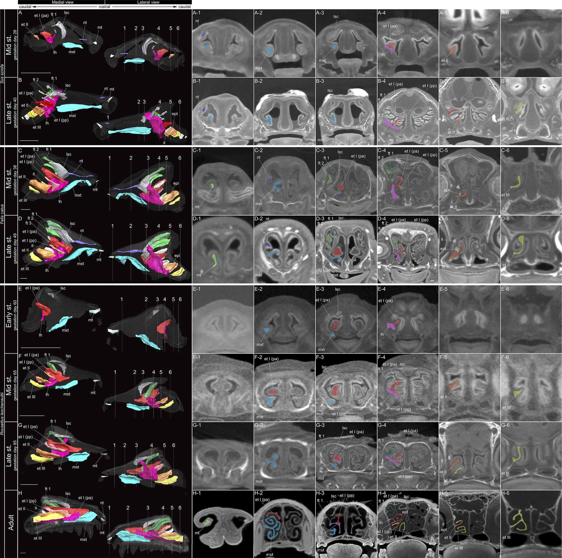
Coronal section and sagittal 3D views of µCT images of Sus scrofa, Felis catus, and Rousettus leschenaultii. A–H Show approximate location of section through the nasal capsule or nasal cavity. (A-1–6) Mid stage fetus and (B-1–6) late stage fetus of Sus scrofa. (C-1–6) Mid stage fetus and (D-1–6) late stage fetus of Felis catus. (E-1–6) Early stage fetus, (F-1–6) mid stage fetus, (G-1–6) late stage fetus, and (H-1–6) adult of Rousettus leschenaultii. Scale bars: 1 mm. Abbreviations: ept = epiturbinal; et I (pa) = ethmoturbinal I pars anterior; et I (pp) = ethmoturbinal I pars posterior; et II–III = ethmoturbinal II–III; ept = epiturbinal; ft 1–3= frontoturbinal 1–3; it = interturbinal; lh = lamina horizontalis; lsc = lamina semicircularis; mt = marginoturbinal; mxt = maxilloturbinal; nt = nasoturbinal. |