|
||
|
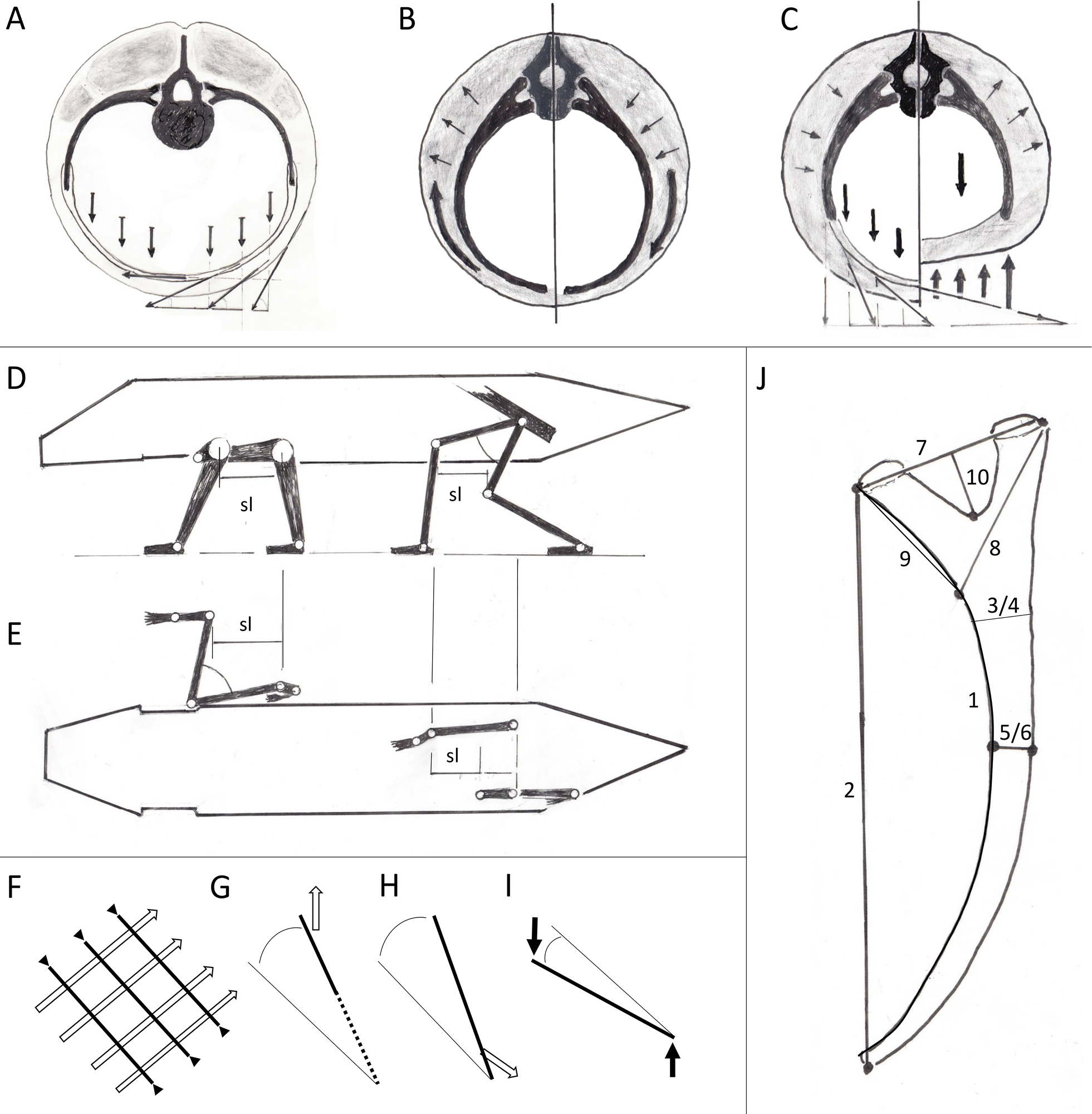
A–C Functional loading of ribs on cross sections. Skeletal elements drawn thicker than musculature. A The weight of the intestines pushes downward and is represented by six arrows. On the right side, the parts of weight combine with the pulling force of the muscles to resultants, which agree with the local direction of the muscles. The ventral tips of the ribs are pulled downward, and can be sustained because of the long, bifurcated collum and tuberculum. B Torsional moments compress the ribs on one side, while extending them on the other. In the first case, ribs tend to vault laterally, in the second, the curvature becomes flatter. C In contrast to the trunk carried freely above the ground (left side, also in A), a belly-dragging posture (right side) leads to compression of the ribs, not to tension. Ribs in side view. D, E Sketch of a synapsid, with sprawling forelimbs and parasagittally moving hindlimbs. D Lateral, E top view. Segment length is the same in both sketches, the excursion of the hip and shoulder joint are identical, as well as the step length sl. The excursion range of the zeugopodium is enabled by a rotation about the long axis of the stylopodium. F Direct transfer of body weight on the ground (bold arrows) leads to a more inclined position of the rib (after Preuschoft et al. 2007 a and b). This arrangement is suited to withstand the torsional stresses. It changes to its opposite at each step. G M. serratus pulls the anterior ribs cranially and hereby reduces their optimal angle of 45° as shown in F. H The muscles of the body wall pull the middle and posterior ribs downward and readjust their angle towards 90°. I Black arrows indicate compressed ribs, white arrows indicate tension of the intercostal muscles. J Rib measurements. We measured where possible due to preservation, the vertebrae and ribs (with calipers and a measureing tape) of Sauroctonus parringtoni (GPIT-PV-31579), Stahleckeria potens (GPIT-PV-30792), Keratocephalus moloch (GPIT-PV-31461), Belesodon magnificus (GPIT-PV-31575), Hyperodapedon sanjuanensis (GPIT-PV-31578), Dimetrodon limbatus (GPIT-PV-31373). We measured the vertebrae as follows: centrum hight (cranial side), width (cranial side), and length of the second last presacral vertebra to approximately half the tail length, if the respective vertebrae were preserved and if the state of preservation allowed it. Rib measurements included: measurements 1–10. |