|
||
|
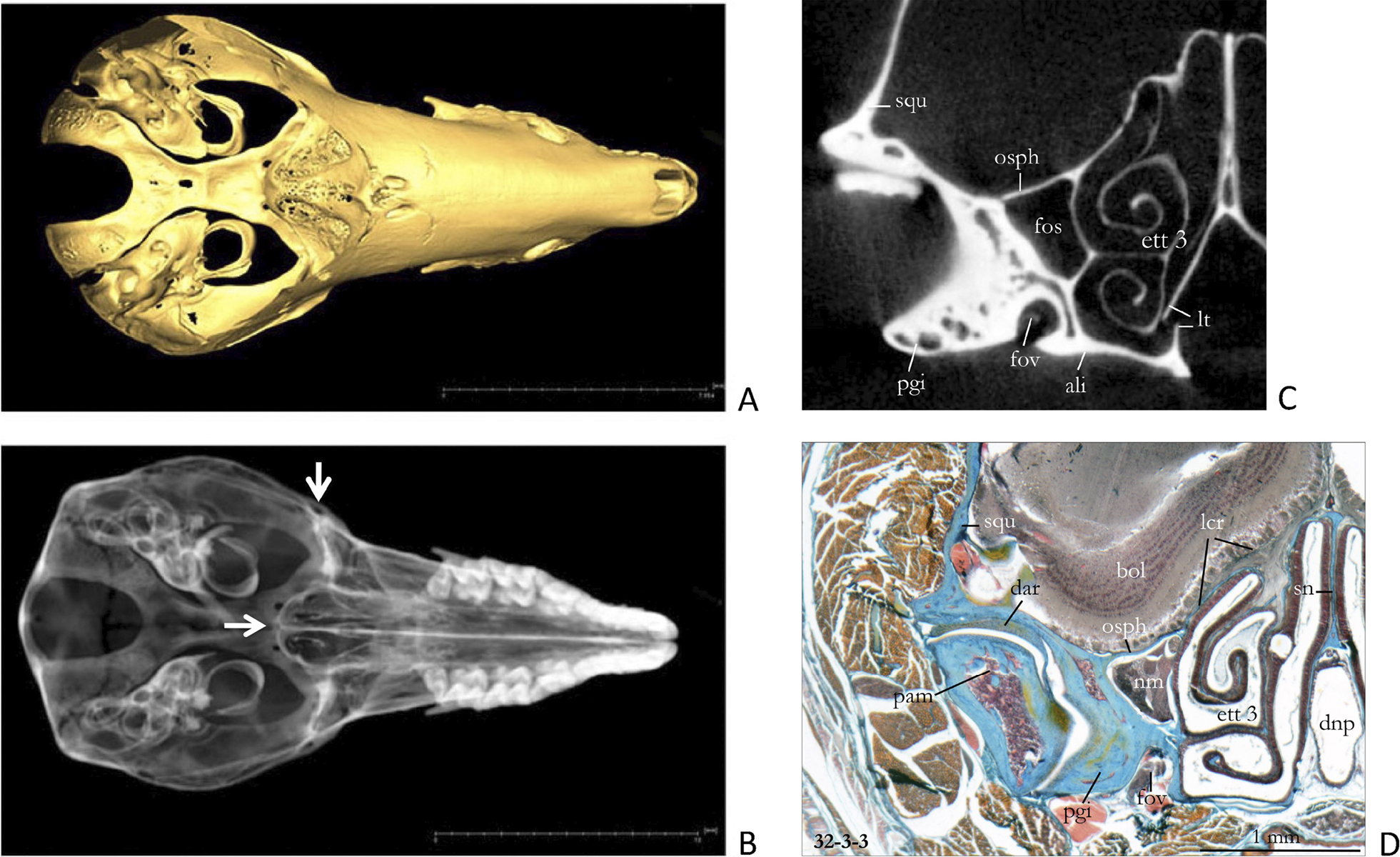
A Virtual picture of the skull of an adult specimen of Sorex araneus (SMF 82598). The roof of the braincase is removed to show the internal cranial base. B This semi-transparent figure (pseudo x-ray) of the same skull shows that the glenoid fossa (vertical arrow) appears to be attached to the posterior part of the nasal capsule. The recessus ethmoturbinalis and the posterior cupula of the nasal capsule reach back to the carotid foramina (horizontal arrow). C Virtual cross section (scan no. 1205, at about the level of the vertical arrow in C) showing the fixation of the glenoid portion of the squamosal to the lateral wall of the nasal capsule. The lamina terminalis is broken in this specimen. D Histological section of an adult specimen of Sorex araneus (Coll. W. Maier) at about the same plane as the µCT scan. d1 32—3-3 means section series of neonate specimen: plate 32, row 3, and section 3. The fissura orbitalis superior is almost completely occupied by the ramus maxillaris of the trigeminal nerve and by the thin nervus opticus in its mediodorsal corner. The anterior notch of the foramen rotundum contains the ramus mandibularis of the trigeminal nerve. Note the pronounced dorsal protrusion of the posterior nasal capsule into the brain cavity and the peculiar structure of the jaw joint. The cross sections C and D are mirrored. Abbreviations: ali – alisphenoid, bol – bulbus olfactorius, dar – discus articularis, dnp – ductus nasopharyngeus, ett 3 – ethmoturbinal 3, fos – fissura orbitalis superior, fov – foramen ovale, lcr – lamina cribrosa, lt – lamina terminalis, nm – nervus maxillaris, osph – orbitosphenoid, pam – processus articularis mandiblae, pgi – processus glenoideus inferior, sn – septum nasi, squ – squamosal. |