|
||
|
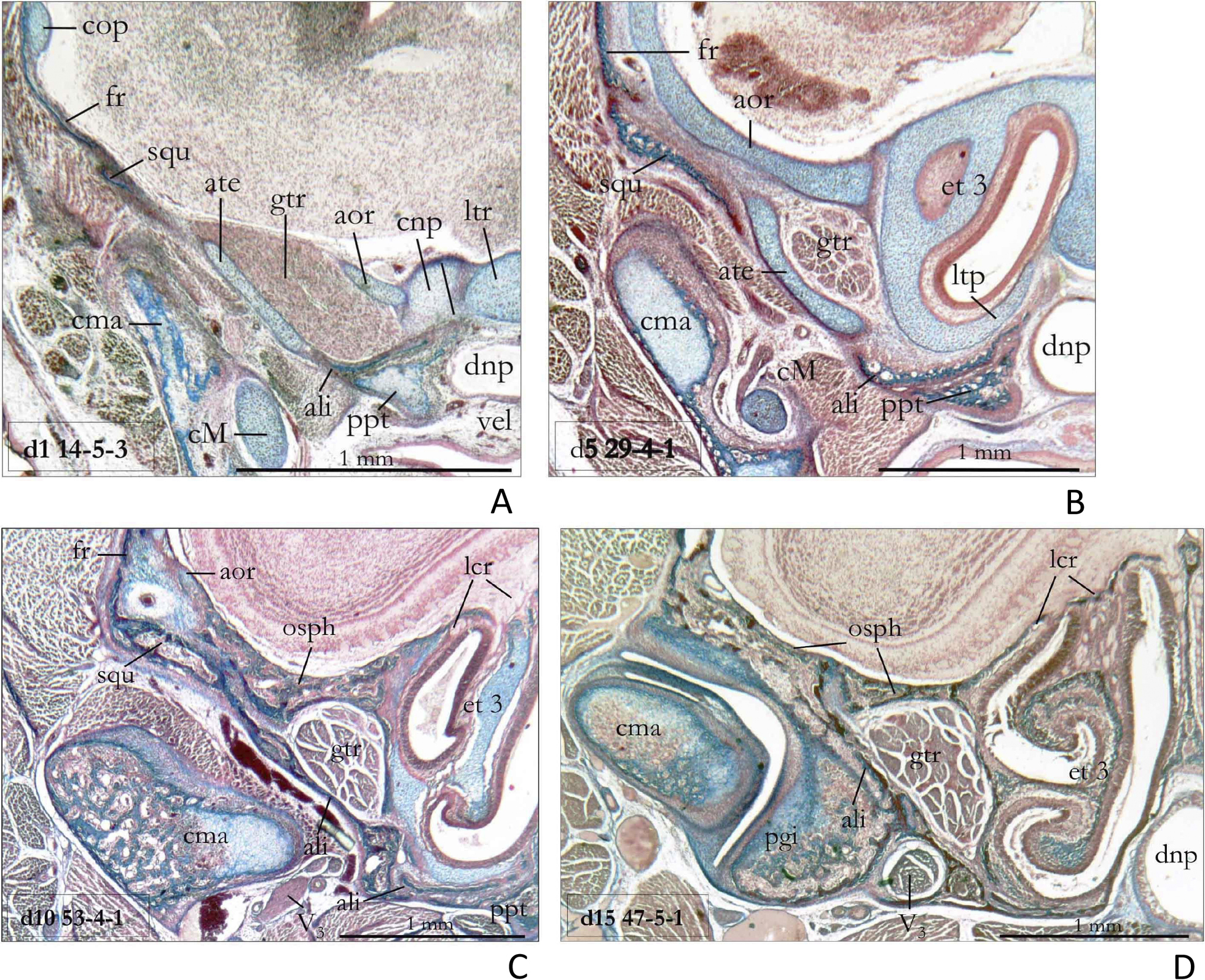
Ala orbitalis, ala temporalis and jaw joint in different postnatal stages of Sorex araneus. A Section d1 14-5-3 shows that the anterior tip of the squamosal does not yet contact the ala orbitalis in the neonate. The anlage of the jaw joint is very immature, and it is still situated at the level of the cupula nasi posterior. B At day 5 (section d5 29-4-1), the squamosal and the ‘anlage’ of the jaw joint have reached the ala orbitalis and both are already positioned at the level of ethmoturbinal 3. However, frontal bone, ala orbitalis, ala temporalis and squamosal are not yet closely connected. C At day 10 (section d10 53-4-1), the orbitosphenoid and the alisphenoid are providing solid supports for the squamosum. D At day 15 (section d15, 47-5-1) we already see similar structural proportions as in the adult (cf. Fig. 1 D). The foramen ovale for the mandibular branch of the trigeminus nerve is situated directly behind the fully developed processus glenoideus inferior. Abbreviations: see Fig. 9, ate – ala temporalis, ltp – lamina transversalis posterior, ltr – lamina trabecularis, pgi – processus glenoideus inferior, V3 – nervus mandibularis, vel – velum palatinum. |