|
||
|
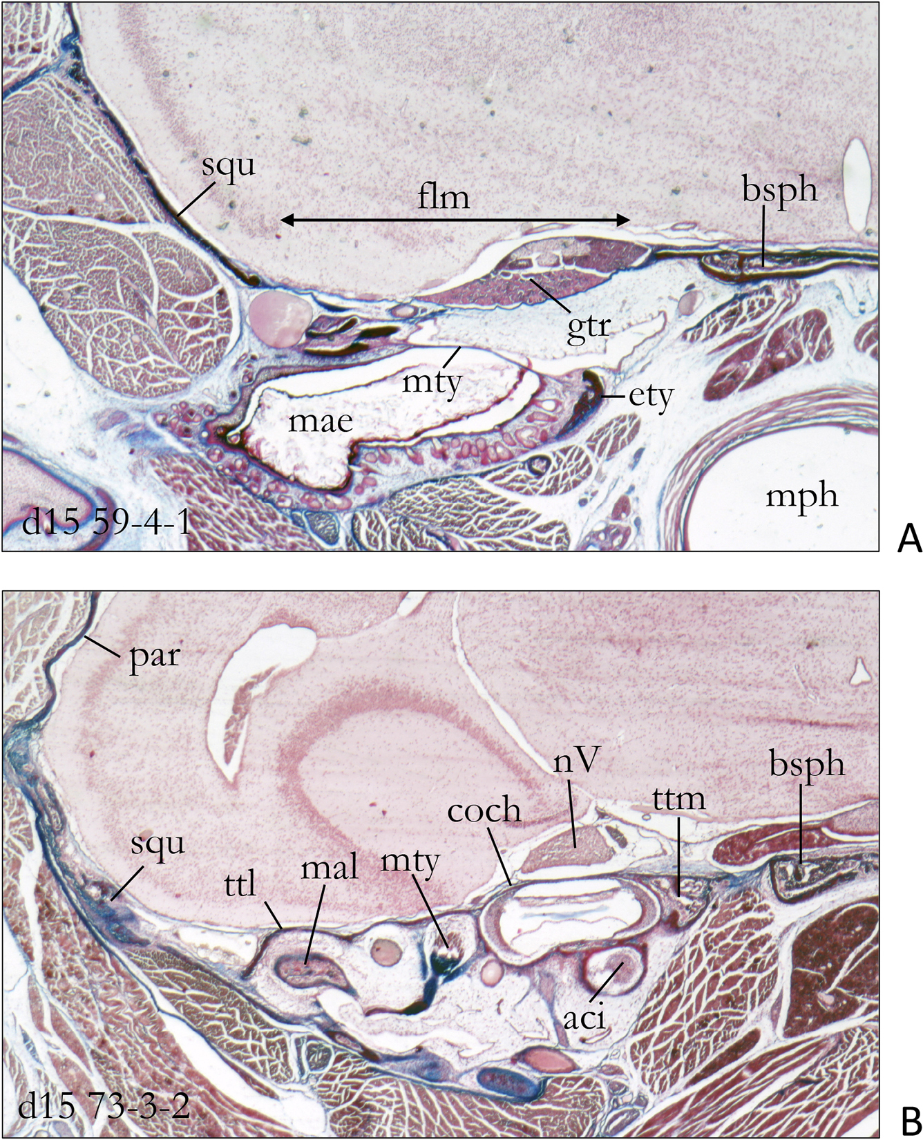
Histological cross sections of the posterior end of the foramen lacerum medium in a two weeks old specimen of Sorex araneus. A In this cross section the foramen lacerum medium spans beween the basisphenoid and the squamosum; it is closed by the posterior sphenobturate membrane. On the dorsal side of the membrane lies closely attached the trigeminal ganglion, i.e. it represents the wall of the cavum epiptericum. Below the membrane we see the rostral parts if the tympanic cavity with the ectotympanic and the tympanic membrane. B More posteriorly the roof of the tympanic cavity is tegmen tympani and the cochlea. Abbreviations: aci – arteria carotis interna, bsph – basisphenoid, coch – cochlea, ety – ectotympanicum, flm – foramen lacerum medium, gtr – ganglion trigemini, mae – meatus acusticus externus, mal – malleus, mph – mesopharynx, mtty – musculus tensor tympani, mty – membrana tympani, nV – nervus trigeminus, par – parietale, squ – squamosum, ttl – tegmen tympani laterale, ttm – tegmen tympani mediale. |