|
||
|
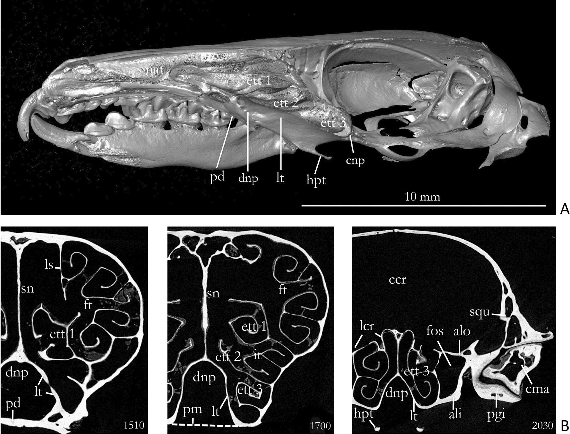
A Paramedian section of a skull of Crocidura russula (Coll. W. Maier). This figure shows the 3 ethmoturbinals filling the recessus ethmoturbinalis. The lamina terminalis is very elongated. B µCT images of the same skull. – Cross section 1510 is near the anterior end of the lamina terminalis. – Section 1700 shows the lamina terminalis in about the midth of the nasopharyngeal duct, which is ventrally closed by the soft palate (stippled line). Cross-section 2030 shows the posterior part of the recessus ethmoturbinalis with the tips of the hamuli pterygoidei. The wall of the braincase and the snout may be interpreted as a strong shell (see discussion). Abbreviations: ali – alisphenoid, alo – ala orbitalis/orbitosphenoid, ccr – cavum cranii, cma – caput mandibulae, cnp – cupula nasi posterior, dnp – ductus nasopharyngeus, ett 1 – ethmoturbinal 1, ett 2 – ethmoturbinal 2, ett 3 – ethmoturbinal 3, fos – fissura orbitalis superior, ft – frontoturbinal, hpt – hamulus pterygoideus, lcr – lamina cribrosa, ls – lamina semicircularis, lt – lamina terminalis, nat – nasoturbinal, pd – palatum durum (secondary palate), pgi – processus glenoideus inferior, pm – palatum molle (velum palatinum), sn – septum nasi, squ – squamosum. |