|
||
|
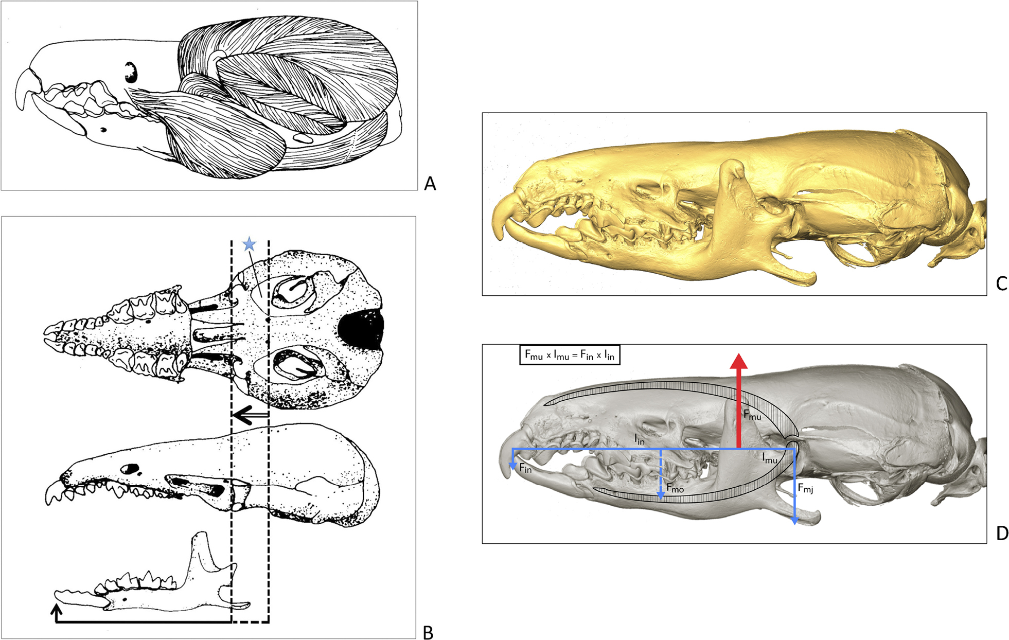
A Complex adductor muscles in Suncus murinus (after Dötsch 1983). B Ventral and lateral view of a skull and mandible of Sorex araneus (after Dötsch 1982). The arrow shows the positional shift of the jaw joint by about 2,2 mm. The solid beam represents the shortened loadlever (– 18 %) for the anteriormost biting point, by which the prominence of the incisors is compensated for. The jaw articulation is detached from the periotic ossification – a unique feature in mammals. The wide fenestra at the cranial base (blue asterisk) is interpreted as foramen lacerum medium (sensu Starck 1979). C Lateral view of a skull of Crocidura russula (Coll. W. Maier no. 3, virtual reconstruction of a µCT scan) with the molars in maximal occlusion. For the occlusion of the edge of the incisors, the mandible must be lowered at the secondary jaw joint. D The same specimen of Crocidura with a simplified drawing of the basic mechanic vectors and trajectories of the upper and lower jaws: Black colour – upper and lower jaws conceived as a pair of tweezers; blue colour – biting forces at the first incisors (Kin) and at the molars (Kmo) and jaw load Kmj. Red colour (Fmu) – estimated resultant of vertical adductor muscle forces with muscle-lever (Lmu). All partial forces were calculated in relation to Fmu = 1. |