|
||
|
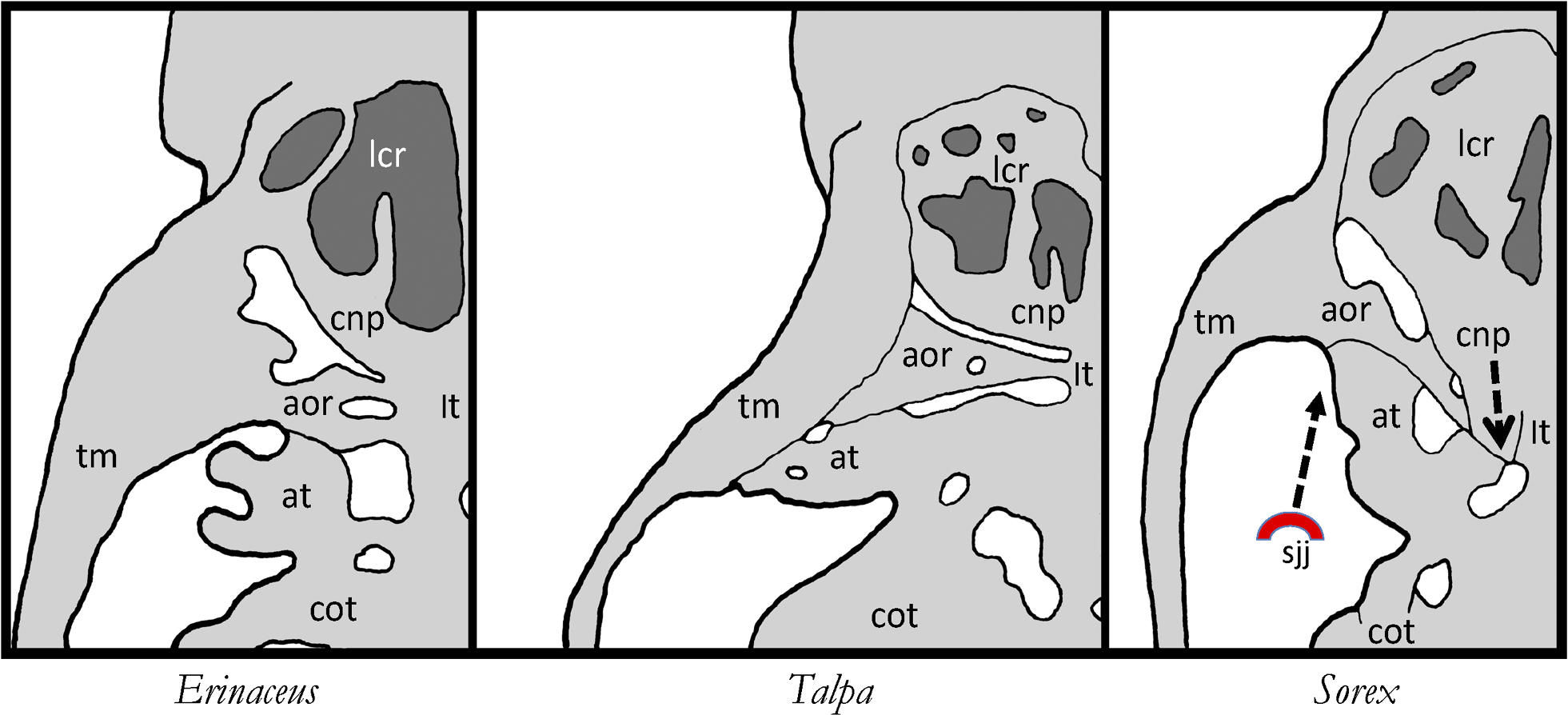
The endocranial base in three fetal Lipotyphla: Erinaceus europaeus (from Fawcett 1918), Talpa europaea (from Fischer 1901) and Sorex araneus (from DeBeer 1929). By the backward expansion of the posterior nasal capsule in Sorex, its ala orbitalis is largely separated from the trabecular plate of the central stem (remnants of this contact is described in the text). The tiny foramen opticum is medially framed by the lateral wall of the nasal capsule. In this fetus of Sorex the ‘anlage’ of the secondary jaw joint (ssj) is still situated at some distance from the ala temporalis and the cupula nasi posterior; therefore, it cannot be primarily causal to the displacement of these structures. Abbreviations: aor – ala orbitalis, at – ala temporalis, cnp – cupula nasi posterior, cot – capsula otica, lcr – lamina cribrosa, lt – lamina trabecularis, sjj – secondary jaw joint, tm – taenia marginalis. |