|
||
|
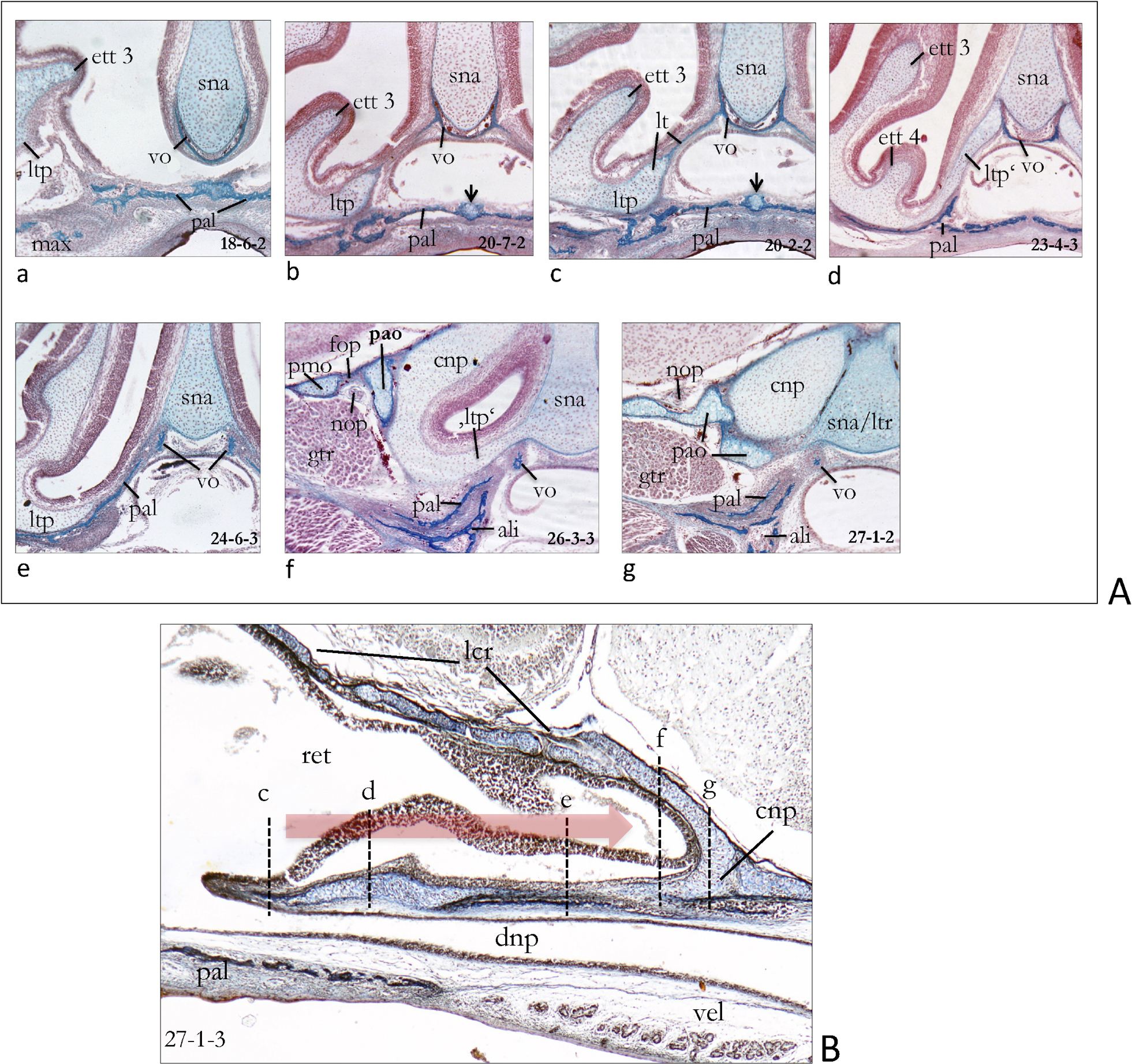
A Lamina terminalis in a neonate Crocidura russula (d1) with selected cross sections. a (section 18-6-2). Cross section slightly anterior to the lamina transversalis posterior (ltp). (The palate is pushed somewhat dorsally and therefore the internasal passage looks too narrow in b and c). b (section 20-7-2). Anterior end of the lamina transversalis posterior showing the heterogeneous origin of the lamina terminalis. Notice the secondary cartilage in the medial palatine suture (arrow). c (section 20-2-2). The complete lamina terminalis is a mixed bone (‘Mischknochen’) of enchondral bone from the nasal capsule and a lateral process of the dermal vomer. d (section 23-4-3). Behind the bony lamina terminalis there is a short gap which is closed by cartilage of the nasal capsule alone. e (section 24-6-3). Further behind this cartilaginous lamina is replaced by the ascending process of the palatine. f (section 26-3-3). Towards the cupula nasi posterior, the lamina transversalis posterior is again cartilaginous. The palatine is underlain by the alisphenoid. g (section 27-1-2). The ventral process of the pila praeoptica is bent around the ventral side of the posterior end of the massive cupula nasi posterior to reach its origin at the nasal septum. B Parasagittal section of a very young postnatal specimen of Crocidura russula (Coll. W. Maier, 21b) showing the recessus ethmoturbinalis and the lamina transversalis posterior. Letters c–f correspond approximately to the cross sections in A. Abbreviations: ali – alisphenoid, cnp – cupula nasi posterior, dnp – ductus nasopharyngeus, ett 3 – ethmoturbinal 3, fop – foramen opticum, gtr – ganglion trigemini, lcr – lamina cribrosa, lt – lamina terminalis, ltr – lamina trabecularis, ltp – lamina transversalis posterior, ltp’ – medial transversal lamina, ‘ltp’ – terminal transversal lamina, max – maxillary, nop – nervus opticus, pal – palatinum, pao – pila praeoptica, fop – foramen opticum, pmo – pila metoptica, ret – recessus ethmoturbinalis, sna – septum nasi, vel – velum palatinum, vo – vomer. |