|
||
|
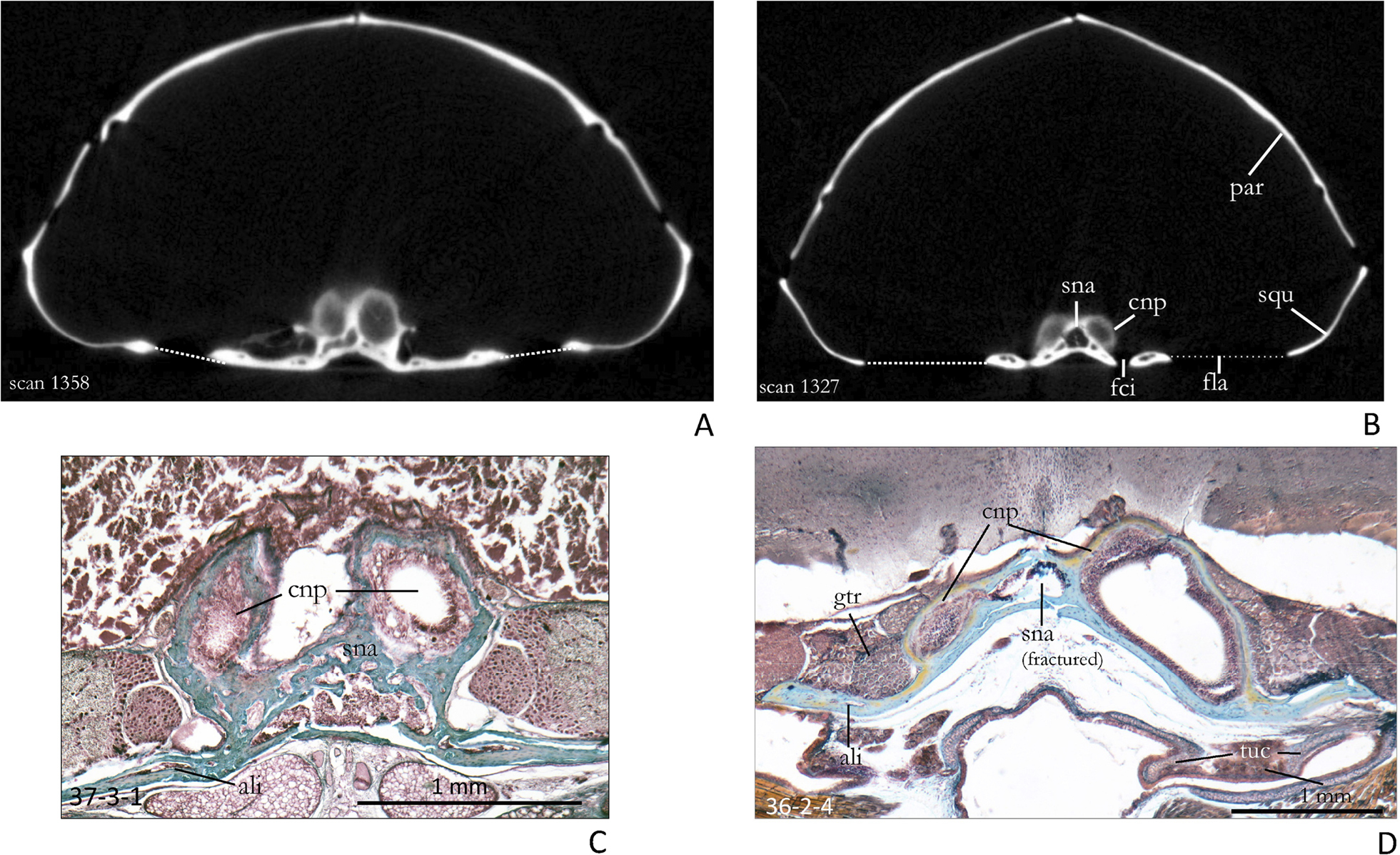
A µCT scan at the level of the cupula nasi posterior in an adult specimen of Crocidura russula (SMF 95044, scan 1358). The septum between the cupulae is narrow. The brain cavity shows quite different proportions in the two taxa. B µCT-scan of an adult specimen of Sorex araneus (SMF 82598, scan 1327). The septum between the cupulae is broad and triangular. The foramen caroticum internum and the anteriormost part of the foramen lacerum medium (stippled line) are also met; they reach further forward in Sorex. The squamosal is more narrow than in Crocidura. C Histological section of the cupula nasi posterior of an adult of Crocidura russula (Coll. W. Maier, section 37-3-1). The ossification of the cupula nasi posterior is still trabecular. D Histological section of an adult specimen of Sorex araneus (Coll. W. Maier, section 36-2-4). All bony elements are fused and transformed into lamellar bone. The posterior septum nasi is broad. The dorsal projection of the cupula nasi posterior of Crocidura is more pronounced than that of Sorex. Abbreviations: ali – alisphenoid, cnp – cupula nasi posterior, fci – foramen caroticum internum, fla – foramen lacerum medium, gtr – ganglion trigemini, par – pariatal, sna – septum nasi (sphenethmoid), squ – squamosal, tuc – cartilago tubae auditivae. |