|
||
|
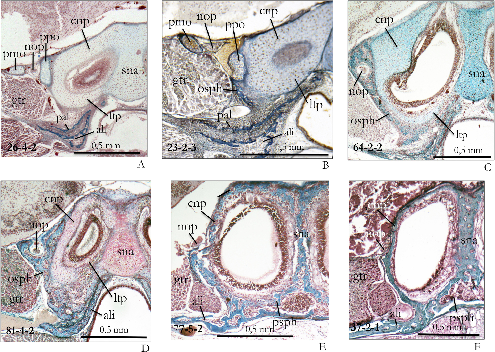
The cupula nasi posterior and orbitosphenoid in different ontogenetic stages of Crocidura russula. – A Neonate specimen (d1, section 26-1-2). The foramen opticum is shifted to the lateral side of the posterior nasal cupula. The large ganglion trigemini occupies almost completely the fissura orbitalis superior. B Young postnatal stage (Coll. W. Maier; section 23-2-3). The pila praeoptica forms ‘Zuwachsknochen’ (orbitosphenoid) at its ventral edge. The pila metoptica is partially ossified. C This specimen was 5 days old (d5, section 64-2-2). The expanded ‘Zuwachsknochen’ of the orbitosphenoid has enclosed the nervus opticus in a foramen opticum and spreads at the ventral side of the cupula nasi posterior. D This specimen was 8 days old (d8, section 81-4-2). The ‘Zuwachsknochen’ has almost completely enclosed the ventral parts of the cartilaginous cupula posterior. The cartilages of the cupula show first signs of reduction. The septum nasi begins to ossify endochondrally. E This specimen was 15 days old (d15, section 77-5-2). The cartilage has completely disappeared – probably resorbed. The narrow septum nasi is completely ossified and the orbitosphenoid ossification is fused with the alisphenoid. F This young adult still shows trabecular bone in the cupula nasi posterior, the nasal septum and the alisphenoid (cf. Fig. 5C). Abbreviations for Figs 6 and 7: ali – alisphenoid, cnp – cupula nasi posterior, fop – foramen opticum, gtr – ganglion trigemini, hpt – hamulus pterygoideus, ltp – lamina transversalis posterior, ltr – lamina trabecularis, nop – nervus opticus, osph – orbitosphenoid (‘Zuwachsknochen’), pal – palatinum, pmo – pila metoptica, ppo – pila praeoptica, ppt – processus pterygoideus, psph – praesphenoid, sna – septum nasi. |