|
||
|
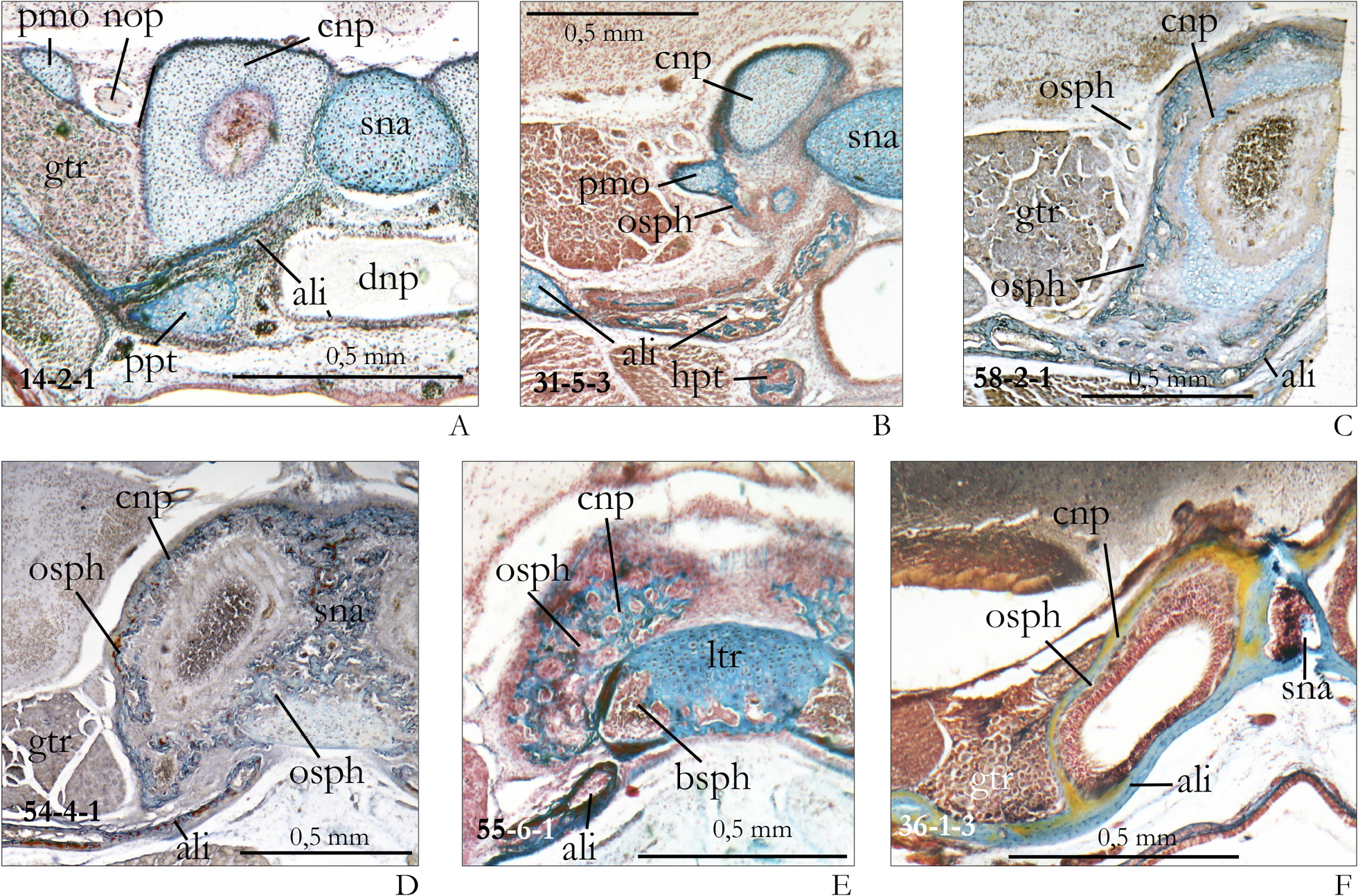
Posterior nasal cupula and orbitosphenoid in different ontogenetic stages of Sorex araneus. A (d1, section 14-2-1). In the neonate of Sorex araneus the cupula nasi posterior is still cartilaginous; the foramen opticum is framed by the pila metoptica only. The ganglion trigemini lies next to the cupula and is ventro-laterally supported by the ala temporalis. B (d5, section 31-5-3). The cupula nasi posterior and the pila metoptica of a Sorex-specimen of 5 days show the initial formation of ‘Zuwachsknochen’. C (d10, section 58-2-1). In this 10 days old specimen, the cartilaginous cupula nasi posterior is almost completely enclosed by Zuwachsknochen (orbitosphenoid) and the cartilage is in the process of resorption or endochondral ossification. D (d15, section 54-4-1). At this stage of 15 days, the cartilage of the cupula nasi posterior has completely disappeared and is replaced by Zuwachsknochen (orbitosphenoid). It is also fused with the bony nasal septum and with the cupula nasi of the other side. E (d15, section 55-6-1 of the same spcimen) shows the very end of the bony cupula nasi. The edges of the trabecular plate are already ossified as basisphenoid. F (adult, section 36-1-3). In this adult specimen the cupula nasi posterior is formed by thin lamellar bone; it is fused with the bony septum nasi and the alisphenoid. Abbreviations: see Fig. 6, bsph – basisphenoid, dnp – ductus nasopharyngeus, nop – nervus opticus. |