|
||
|
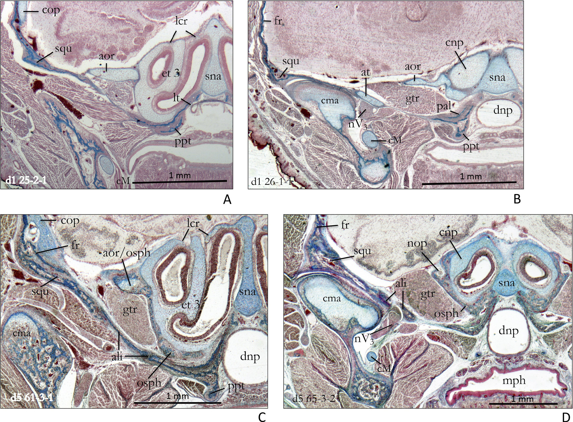
Ala orbitalis and squamosum in a neonate (above) and a five days old specimen (below) of Crocidura russula. A In section d1 25-2-1 the cartilaginous ala orbitalis has only feeble contact with the rostral end of the squamosum. B Section d1 26-1-1 shows that at the level of the initially differentiated jaw joint the squamosum is in contact with the ala temporalis. The proximal edge of the ala orbitalis is connected with the trabecular plate by the rudimentary pila metoptica. C At d5 61-3-1 the ala orbitalis is ossified in its distal and proximal parts, and the pila metoptica is partly developed into an expanded orbitosphenoid by ‘Zuwachsknochen’. D In section d5 65-3-2 the bony orbitosphenoid has almost completely enclosed the cartilaginous cupula nasi posterior. The glenoid region of the squamosum is closely connected with the alisphenoid. The lateral and posterior part of the alisphenoid is pierced by the foramen ovale for the ramus mandibularis of the nervus trigeminus. Abbreviations: ali – alisphenoid, aor – ala orbitalis, at – ala temporalis, cM – cartilago Meckeli, cma – caput mandibulae, cnp – cupula nasi posterior, cop – commissura orbitoparietalis, dnp – ductus nasopharyngeus, et 3 – ethmoturbinal 3, fr – frontale, gtr – ganglion trigemini, lcr – lamina cribrosa, lt – lamina terminalis, mph – mesopharynx, nop – nervus opticus, nV3 – nervus mandibularis, osph – orbitosphenoid, ppt – processus pterygoideus, sna – septum nasi, squ – squamosum. |